
 By admin
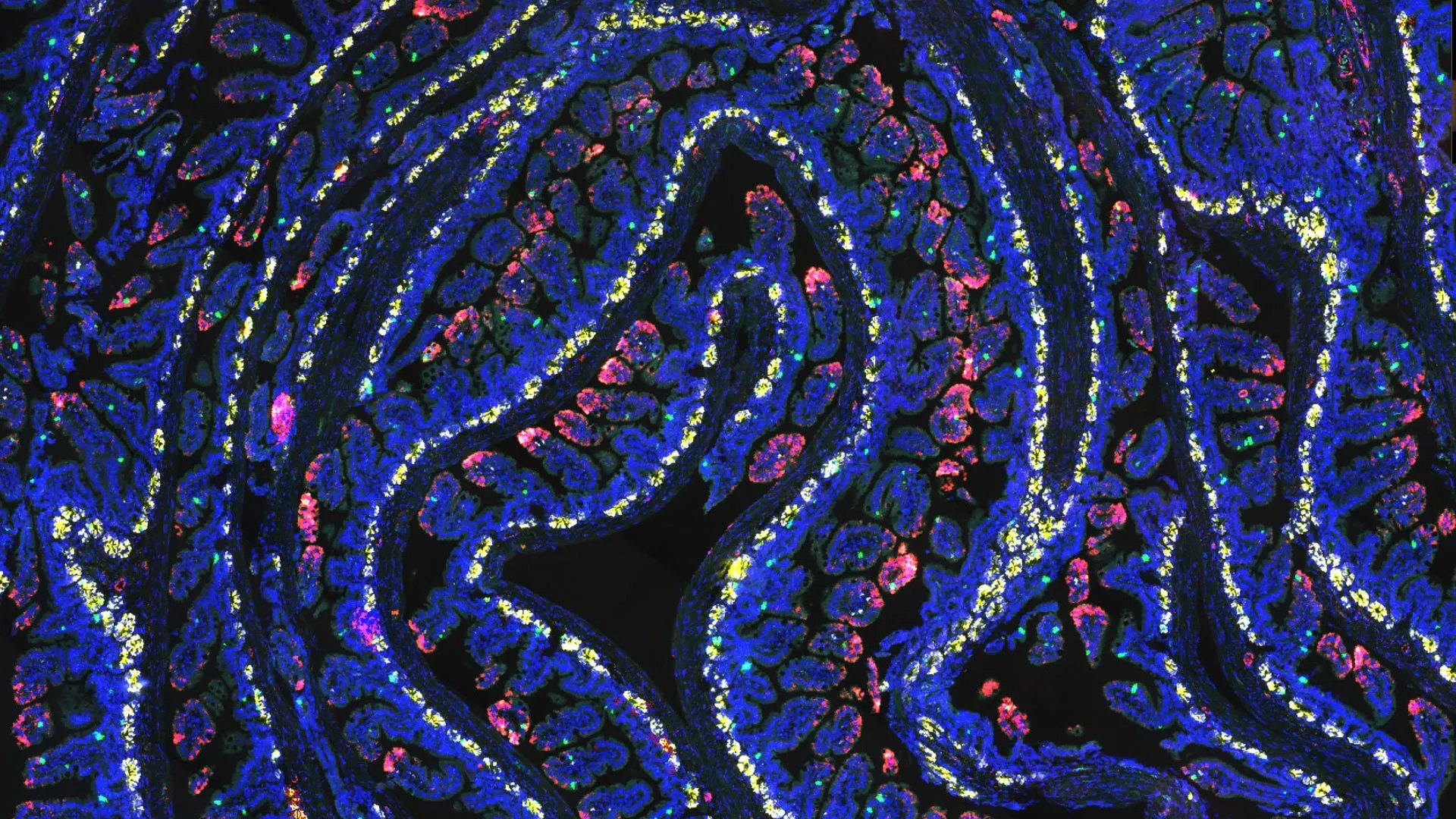
This pivotal discovery could profoundly reshape the landscape of supportive care in oncology, eventually leading to innovative strategies to protect healthy tissue during cancer treatment. By bolstering the intestine’s innate ability to recover from radiation exposure, researchers harbor significant hope of mitigating severe, often debilitating side effects, thereby enhancing patient quality of life and potentially rendering radiation therapy both safer and more efficacious. The implications extend beyond just comfort, touching upon the very feasibility and intensity of life-saving treatments.
The comprehensive findings, which mark a significant leap forward in understanding host-microbe interactions in cancer care, were meticulously documented and published in the prestigious Proceedings of the National Academy of Sciences. The groundbreaking study was collaboratively spearheaded by two distinguished leaders in their fields: Helen Piwnica-Worms, Ph.D., a revered professor of Experimental Radiation Oncology, and Kunal Rai, Ph.D., an eminent professor of Genomic Medicine, both affiliated with the world-renowned MD Anderson Cancer Center. Their combined expertise in radiation biology and genomics proved instrumental in unraveling this complex biological puzzle.
Dr. Piwnica-Worms eloquently articulated the essence of their findings, stating, "Fasting helps prepare intestinal cells to respond more quickly and effectively after injury, almost like training the cells with an emergency preparedness plan." She continued, "This study helps explain how that plan is organized at a molecular level and, crucially, identifies a key bacterium involved in coordinating this sophisticated, protective response." Her analogy underscores the proactive cellular conditioning that fasting facilitates, transforming a potentially vulnerable system into a resilient one.
Understanding the Vulnerability: Why Radiation Therapy Damages the Intestines
Radiation therapy stands as a cornerstone in cancer treatment, globally employed in over half of all cancer cases. It is particularly indispensable for targeting cancers located in the abdomen and pelvis, including highly prevalent and aggressive forms such as pancreatic, colorectal, gynecologic (ovarian, cervical, uterine), and prostate cancers. While undeniably effective in its primary mission of annihilating rapidly dividing cancer cells by damaging their DNA, radiation therapy operates with a certain degree of collateral damage. The energy delivered by radiation beams, though precisely aimed, inevitably interacts with surrounding healthy tissues, leading to undesirable side effects.
The small intestine, a vital organ responsible for nutrient absorption, is notoriously vulnerable to this collateral harm. Its delicate lining, known as the intestinal epithelium, is characterized by an exceptionally high cellular turnover rate, making its rapidly dividing cells exquisitely sensitive to radiation exposure. This sensitivity stems from the fact that radiation targets cells in their proliferative phases, which are abundant in the intestinal crypts where stem cells constantly regenerate the epithelial lining. When this protective and absorptive lining is compromised, patients frequently endure a spectrum of distressing symptoms including severe nausea, debilitating diarrhea, abdominal pain, and an increased susceptibility to opportunistic infections due to a compromised gut barrier. In severe cases, radiation-induced enteritis can lead to life-threatening complications such as intestinal perforation, strictures, fistulas, and chronic malabsorption, necessitating surgical intervention and significantly impacting long-term quality of life. These grave risks often compel oncologists to limit the total radiation dose or interrupt treatment schedules, potentially undermining therapeutic efficacy and restricting viable treatment options for patients. Current supportive care strategies, primarily symptomatic (e.g., anti-diarrheals, antiemetics), offer limited proactive protection against the fundamental cellular damage.
Previous preclinical research conducted within the esteemed Piwnica-Worms Laboratory had already hinted at the protective power of dietary intervention. Their earlier studies demonstrated that a period of fasting prior to radiation exposure significantly aided the intestines in their subsequent recovery. This initial observation was highly intriguing, but the precise molecular and cellular mechanisms underpinning this remarkable protective effect remained elusive, representing a significant gap in scientific understanding. The "why" behind the observed benefits was the next frontier.
The latest study, building upon this foundational work, provides a critical piece of that intricate puzzle. It not only identifies a specific, hitherto underappreciated gut bacterium but also meticulously delineates the sophisticated molecular pathways through which it collaborates with fasting to prime intestinal cells for injury and subsequent repair. This deeper understanding moves the field closer to actionable interventions.
The Orchestration of Recovery: How a 24-Hour Fast Activates Gut Repair
The researchers’ investigation unveiled a fascinating cascade of events triggered by a mere 24 hours of fasting. Their findings indicated that this brief period of nutrient restriction dramatically increased the abundance of a beneficial commensal bacterium known as Akkermansia muciniphila, or AKK, specifically within the small intestine. Akkermansia muciniphila is a prominent inhabitant of the human gut, celebrated for its role in maintaining gut barrier integrity by degrading mucin (the primary component of the protective mucus layer) and its association with various positive metabolic health outcomes, including improved glucose metabolism and reduced inflammation. Its increased presence during fasting was a critical initial observation.
Crucially, the study revealed that AKK produces a short-chain fatty acid (SCFA) called propionate. Short-chain fatty acids are metabolic byproducts of bacterial fermentation of dietary fibers in the gut, known for their multifaceted beneficial effects on host physiology, including providing energy for colonocytes and mediating anti-inflammatory responses. In this context, propionate emerged as a key signaling molecule. It was found to work synergistically with other metabolic shifts induced by fasting to initiate profound changes inside intestinal cells, making them remarkably better equipped to regenerate and repair themselves following radiation-induced damage.
The core of this cellular reprogramming lies in the modification of proteins known as histones. Histones are fundamental structural proteins around which DNA is tightly packaged within the cell nucleus, forming nucleosomes. This packaging plays a crucial role in regulating gene expression. The research elucidated that propionate, along with other fasting-induced metabolic alterations, orchestrates specific chemical modifications (e.g., acetylation) to these histones. These epigenetic modifications, which do not alter the underlying DNA sequence but rather influence how genes are read, effectively loosen the DNA’s grip on the histones. This "opening up" of the chromatin structure makes specific portions of the DNA more accessible to the cellular machinery responsible for gene transcription, thereby allowing the cells to activate a specialized repertoire of genes involved in tissue regeneration and repair. During the fasting period, a distinct population of intestinal cells, enriched with these activated repair programs, becomes significantly more abundant and primed for action.
In essence, the 24-hour fast, facilitated by the increased presence and metabolic activity of Akkermansia muciniphila, acts as a sophisticated pre-conditioning regimen. It prepares these intestinal cells by arming them with the precise biological instructions and molecular machinery they will desperately need if an injury, such as radiation exposure, occurs. When the intestines are subsequently exposed to therapeutic radiation, these pre-programmed and prepared cells are able to rapidly expand, proliferate, and differentiate, thereby efficiently rebuilding the damaged intestinal lining and accelerating recovery. This represents a paradigm shift from reactive treatment to proactive cellular preparedness.
The Indispensable Role of One Gut Bacterium in Recovery
To unequivocally ascertain the critical importance of Akkermansia muciniphila in this protective mechanism, the researchers employed rigorous experimental designs. They selectively depleted or removed the bacterium from their preclinical models. The results were striking: without the presence of AKK, the fasting regimen no longer conferred the same profound regenerative and protective benefits to the intestinal tissue. The ’emergency preparedness plan’ seemed to crumble without its key coordinator.
Even more compellingly, the protective response was not only abrogated but could be precisely restored only when the bacterium was reintroduced in combination with fasting. This elegant experimental manipulation definitively demonstrated that neither component alone—neither fasting nor AKK by itself—was sufficient to reproduce the full, robust protective and regenerative effect observed in the study. This synergistic relationship highlights a sophisticated co-dependency, where the metabolic state induced by fasting creates an environment conducive for AKK to thrive and produce its beneficial metabolites, which in turn act on host cells.
The findings thus strongly suggest that fasting achieves more than just a temporary alteration in the gut’s metabolic state. Together with the critical involvement of Akkermansia muciniphila, it fundamentally alters the epigenetic and transcriptional activity of intestinal cells prior to radiation exposure, effectively placing them in a highly optimized state that supports more efficient, rapid, and robust recovery once damage inevitably occurs. This pre-emptive cellular programming represents a novel therapeutic avenue.
By meticulously uncovering this intricate and previously unknown connection between host nutrition, the gut microbiota, and intrinsic cellular regeneration pathways, the researchers at MD Anderson have identified a potential new paradigm for strengthening the body’s resilience to tissue injury. This profound discovery could prove especially valuable for protecting highly sensitive healthy organs and tissues during the aggressive and often damaging regimens of cancer treatment, promising a future where therapeutic efficacy is not inextricably linked to severe systemic toxicity.
Translating Preclinical Promise: Could Gut Bacteria Make Cancer Treatment Safer?
While the results emanating from this preclinical research are unequivocally promising and open exciting new avenues, it is imperative to acknowledge that the research currently resides at the preclinical stage. Significant further investigation will be indispensable to conclusively determine whether the same intricate protective mechanism operates consistently and effectively in human physiology. Furthermore, extensive clinical trials will be required to ascertain whether this mechanism can be safely and reproducibly harnessed to improve clinical outcomes for patients undergoing radiation therapy.
The potential implications of this discovery are vast and extend beyond the intestinal lining. One particularly compelling possibility is that the newly elucidated pathway could be leveraged to assist other rapidly dividing healthy tissues in recovering from the debilitating effects of various cancer treatments. Researchers are keenly interested in exploring its applicability to tissues such as bone marrow, which is highly susceptible to damage from chemotherapy and radiation, leading to myelosuppression and severe immune compromise. Other potential targets include the oral mucosa, often afflicted by painful mucositis, and the skin, which can suffer from radiation dermatitis.
If subsequent investigations in human cohorts robustly support these preclinical findings, the pathway to clinical translation would involve carefully designed clinical trials. These trials could rigorously explore whether activating this repair mechanism—either through controlled fasting, targeted microbial interventions, or metabolite administration—confers significant clinical benefits to patients undergoing abdominal or pelvic radiation therapy. Such benefits could manifest as reduced incidence and severity of acute and chronic gastrointestinal toxicities, improved quality of life, fewer treatment interruptions, and potentially the ability to deliver higher, more effective radiation doses.
Crucially, the discovery also illuminates potential pathways to achieve these beneficial effects without necessarily requiring cancer patients to undergo fasting, which can be challenging, impractical, and even detrimental for individuals already struggling with cancer-related cachexia or nutritional deficiencies. This opens the door to developing innovative, non-dietary interventions. For instance, targeted treatments involving the direct administration of beneficial bacteria like Akkermansia muciniphila (as a probiotic) or the precise molecules they produce (such as propionate, as a postbiotic) could potentially stimulate similar repair processes. Such approaches would bypass the practical difficulties and potential risks associated with food restriction during the demanding period of cancer care, offering a more patient-friendly and scalable solution.
Dr. Rai underscored this pragmatic perspective, stating, "Fasting is not always practical or advisable for cancer patients, who often face significant nutritional challenges and disease-related weight loss. This foundational work supports several other potential ways to enhance recovery after treatment without necessarily imposing dietary restrictions." He elaborated, "Whether through carefully designed dietary interventions that promote beneficial microbes, the direct administration of targeted microbes themselves, or the therapeutic delivery of their key metabolites, the ultimate goal remains the same: to help repair healthy tissue more effectively and sustainably while patients receive the life-saving cancer therapies they so desperately need." This holistic approach reflects a commitment to improving both survival and quality of life.
This pivotal research was made possible through the generous support and funding from leading national and state scientific organizations, including the National Cancer Institute (NCI) within the National Institutes of Health (NIH), a primary federal agency responsible for cancer research, and the Cancer Prevention and Research Institute of Texas (CPRIT), a state-funded initiative dedicated to advancing cancer research and prevention. Their investment in fundamental research continues to yield discoveries that promise to redefine cancer care.
