
Now, a groundbreaking development from the Korbel Group at the European Molecular Biology Laboratory (EMBL) Heidelberg offers unprecedented insight into this fundamental aspect of cancer initiation. Researchers there have engineered a powerful, artificial intelligence-driven platform designed to systematically investigate the precise conditions and mechanisms that lead to the formation of these critical chromosomal abnormalities. By meticulously dissecting the molecular milieu and cellular events that foster these errors, this innovative technology promises to deepen our understanding of the very genesis of cancer, potentially paving the way for novel diagnostic tools and therapeutic interventions.
Jan Korbel, a Senior Scientist at EMBL and the senior author of the pivotal new paper published in the prestigious journal Nature, underscored the gravity of these genetic aberrations. "Chromosomal abnormalities are a main driver for particularly aggressive cancers, and they’re highly linked to patient death, metastasis, recurrence, chemotherapy resistance, and fast tumor onset," Korbel stated. His team’s primary motivation was to unravel the underlying determinants influencing the likelihood of cells acquiring such profound chromosomal alterations. Furthermore, they sought to quantify the spontaneous rate at which these abnormalities emerge during the division of an ostensibly normal cell, a critical piece of information previously obscured by technical limitations. Understanding these rates and triggers is paramount for deciphering cancer’s initial stages and identifying vulnerable cells.
The profound link between abnormal chromosomes and the development of cancer is not a new concept; it has intrigued scientists for over a century. The German zoologist and cytologist Theodor Boveri first articulated this visionary hypothesis in the early twentieth century. Through his meticulous observations of sea urchin eggs under the microscope, Boveri noticed that abnormal chromosome numbers (aneuploidy) and aberrant mitotic divisions were correlated with abnormal development. He proposed that an incorrect chromosomal content within cells could fundamentally alter their behavior, leading to uncontrolled growth and, ultimately, cancer. Boveri’s "somatic mutation theory" was decades ahead of its time, laying a conceptual foundation that continues to influence cancer research today.
Despite Boveri’s enduring theory, systematically studying these elusive abnormalities has remained an immense challenge. The primary hurdle lies in their transient nature and rarity. At any given moment, only a minuscule fraction of cells within a tissue may exhibit chromosomal defects. Moreover, cells that acquire such severe genomic damage are often naturally culled by the body’s sophisticated surveillance mechanisms, undergoing programmed cell death (apoptosis) or entering a state of irreversible growth arrest (senescence). This natural cellular selection, while protective, makes it exceedingly difficult for researchers to isolate and study these aberrant cells. Traditionally, scientists had to embark on the laborious and time-consuming task of manually scanning countless cells under a microscope, a process that yielded only a handful of candidates for further investigation, severely limiting the scope and scale of studies.
Recognizing these formidable technical barriers, Marco Cosenza, a Research Scientist within the Korbel Group, embarked on a mission to devise a more efficient solution. His journey began through collaborations with other EMBL teams grappling with similar limitations in high-throughput biological imaging and genomic analysis. The synergistic efforts led to the conceptualization and development of an automated platform that seamlessly integrates advanced microscopy, cutting-edge single-cell sequencing technologies, and sophisticated artificial intelligence algorithms. This innovative system, christened machine learning-assisted genomics and imaging convergence, or MAGIC, represents a significant leap forward in cellular anomaly detection.
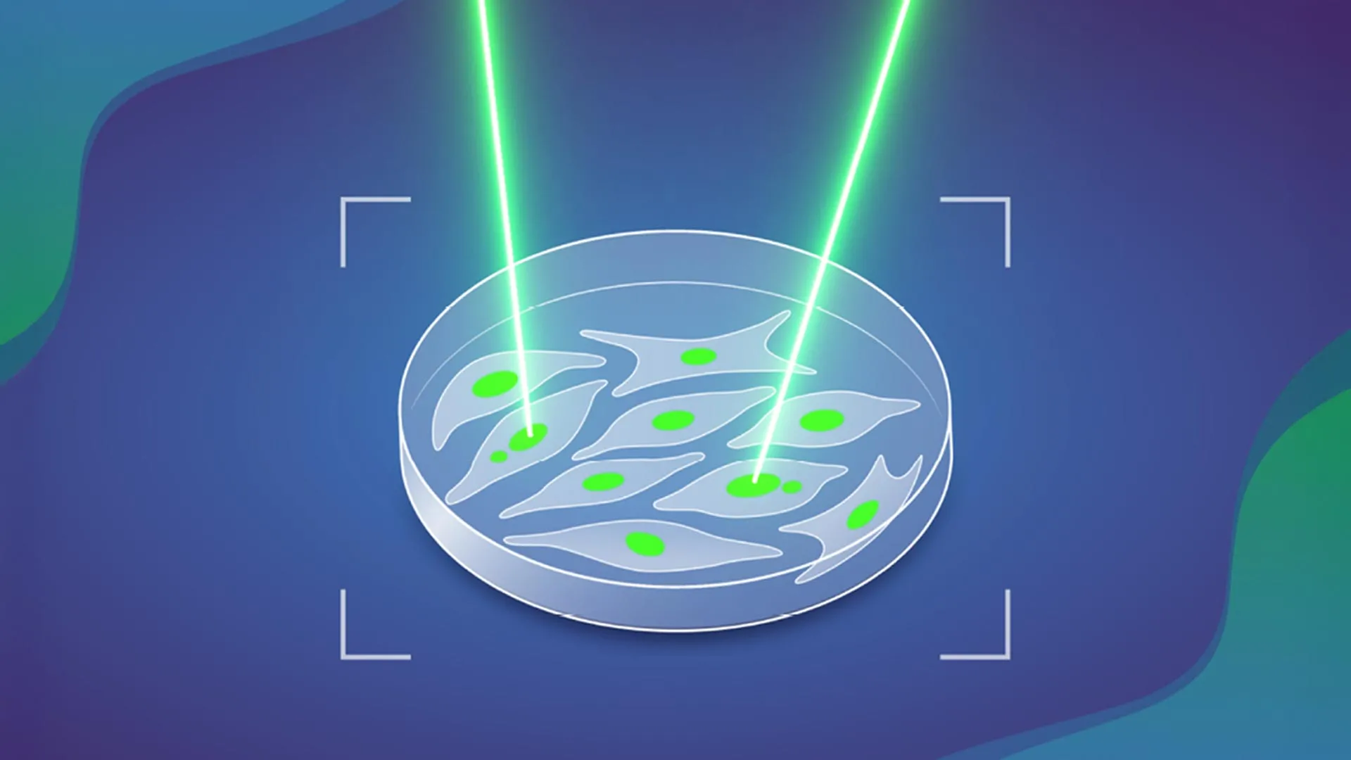
MAGIC operates with remarkable precision, akin to a highly automated version of a "laser tag" game played at the cellular level. The system’s initial step involves rapidly scanning vast populations of cells to pinpoint those displaying a specific, visually discernible feature. In this foundational study, the researchers specifically focused on identifying a distinctive cellular structure known as a ‘micronucleus.’ These small, membrane-bound compartments are often found within the cytoplasm of cells, separate from the main nucleus. They contain fragments of DNA—or even entire chromosomes—that were either lost or improperly segregated during a preceding cell division.
Micronuclei are not merely curiosities; they are critical biomarkers of genomic instability. Cells harboring micronuclei are highly susceptible to undergoing further, more catastrophic chromosomal abnormalities, including events like chromothripsis—a phenomenon where a single chromosome, or a few chromosomes, shatters into many pieces and then reassembles in a scrambled, rearranged fashion. This cascade of genomic damage significantly elevates a cell’s propensity to become cancerous. Therefore, detecting micronuclei serves as an early and crucial indicator of a cell’s increased risk for malignant transformation.
Once the MAGIC system successfully detects a cell containing a micronucleus, it precisely marks that cell using a finely directed laser beam. This ingenious tagging process leverages the properties of a photoconvertible dye—a specialized fluorescent molecule. These dyes exhibit a unique characteristic: upon exposure to light of a specific wavelength, they undergo a chemical transformation that alters the color of the light they emit, or significantly increases their fluorescence intensity. This permanent "tag" effectively labels the cell for subsequent isolation and detailed analysis, a feat impossible with transient markers.
Marco Cosenza reflected on the interdisciplinary nature of the project: "This project combined a lot of my interests in one. It involves genomics, microscopic imaging, and robotic automation." He further highlighted how the unexpected circumstances of the COVID-19-related lockdown in 2020 provided a unique opportunity: "During the COVID-19-related lockdown in 2020, I could really spend some time on learning and applying AI computer vision technologies to the biological image data we had collected before. Afterwards, we designed experiments to validate it and take it further." This period of focused development allowed the team to refine the AI algorithms, transforming raw image data into actionable biological insights.
The MAGIC system orchestrates its complex tasks through a series of seamlessly automated steps. Initially, an automated microscope, equipped with high-throughput imaging capabilities, captures a vast collection of images from a sample of cultured cells. These images are then fed into a sophisticated machine learning algorithm. This algorithm has been rigorously trained using a meticulously curated dataset of manually labeled examples of micronuclei-containing cells, allowing it to accurately recognize the subtle morphological cues indicative of these structures. The training process involves showing the algorithm thousands of images, each annotated by human experts, enabling it to learn and generalize patterns of micronuclei presence.
Should the algorithm successfully identify a cell containing a micronucleus, it instantly relays the precise spatial coordinates of that cell back to the microscope. The microscope then, with pinpoint accuracy, directs a beam of light at the designated cell, activating the photoconvertible dye and permanently tagging it. This "digital-to-physical" feedback loop is the core innovation enabling targeted labeling. Following this tagging, researchers can efficiently isolate these labeled cells from the larger, heterogeneous population of living cells. This is typically achieved using techniques such as flow cytometry (fluorescence-activated cell sorting, or FACS), which employs lasers and detectors to sort cells based on their fluorescent properties. Once isolated, these enriched populations of "at-risk" cells can be subjected to much more detailed and comprehensive study, including in-depth analysis of their genomes, transcriptomes, and epigenomes, providing an unprecedented window into the molecular events driving genomic instability.
By automating what was once an excruciatingly slow and labor-intensive manual search, MAGIC has fundamentally transformed the scale of cellular analysis. The system allows scientists to examine orders of magnitude more cells than was previously conceivable. In a mere matter of hours—less than a single day—MAGIC can process and analyze close to 100,000 cells, generating a wealth of data that would have taken months or even years to acquire through traditional methods. This dramatic increase in throughput is crucial for studying rare cellular events and for performing statistically robust analyses.
Leveraging the power of MAGIC, the researchers embarked on a comprehensive study of chromosomal abnormalities in cultured cells derived from normal human tissues. Their meticulously conducted analysis yielded a striking revelation: slightly more than 10% of spontaneous cell divisions, even in seemingly healthy cells, produce some form of chromosomal abnormality. This baseline rate provides critical context for understanding the inherent genomic instability present in human cells. Furthermore, the team investigated the impact of mutations in the p53 gene, a well-known tumor suppressor often referred to as the "guardian of the genome" due to its critical role in DNA repair and apoptosis. They discovered that when the p53 gene is mutated, the rate of spontaneous chromosomal abnormalities nearly doubles, underscoring the profound protective role of p53 and how its inactivation accelerates genomic chaos, a hallmark of many aggressive cancers.
Beyond p53, the team also delved into other factors influencing the formation of these abnormalities. Their investigations included exploring the presence and specific genomic location of double-stranded DNA breaks within chromosomes. These breaks are among the most deleterious forms of DNA damage, and their faulty repair can directly lead to chromosomal rearrangements and instability. The position of these breaks, particularly within regions known as "fragile sites," was also examined, as these areas are predisposed to breakage and rearrangement.
The success of this ambitious research project was a testament to the power of interdisciplinary collaboration, involving expertise both within and outside EMBL. Key contributors from within EMBL Heidelberg included the Advanced Light Microscopy Facility (ALMF), providing state-of-the-art imaging capabilities, and the Pepperkok Team, renowned for their expertise in quantitative microscopy and cell biology. External collaborations were equally vital, including Isidro Cortes-Ciriano’s group at EMBL-EBI, which brought deep knowledge in computational genomics and artificial intelligence, and Andreas Kulozik’s team at the German Cancer Research Centre (DKFZ). Kulozik’s group, also part of the Molecular Medicine Partnership Unit (MMPU) between EMBL and the University of Heidelberg, provided crucial clinical and translational insights, bridging basic research with potential applications in pediatric oncology.
Crucially, MAGIC has been designed with inherent flexibility and adaptability in mind. While this inaugural study successfully trained the AI to detect micronuclei—a potent indicator of genomic instability—the underlying artificial intelligence architecture is highly versatile. It can be retrained to identify a vast array of other cellular features or anomalies. As Jan Korbel emphasized, "As long as you have a feature that can be discriminated visually from a ‘regular’ cell, you can—thanks to AI—train the system to detect it." This profound adaptability means that MAGIC’s potential applications extend far beyond cancer research. It could be leveraged to study other cellular pathologies, infectious diseases, developmental biology, drug screening, and fundamental processes like cellular senescence or differentiation. "Our system therefore has potential to advance future discoveries in numerous areas of biology," Korbel concluded, hinting at a future where AI-powered microscopy and genomics become indispensable tools for accelerating biological discovery across the scientific spectrum. This development marks a significant stride in our ability to probe the cellular landscape with unprecedented precision and throughput, opening new frontiers in understanding health and disease.