
The pivotal research, conducted by a dedicated team at Oregon Health & Science University (OHSU), was recently published in the esteemed journal Science Advances. This publication brings to light intricate details of the disease’s mechanism, offering a beacon of hope for developing targeted interventions that could fundamentally change the lives of affected individuals.
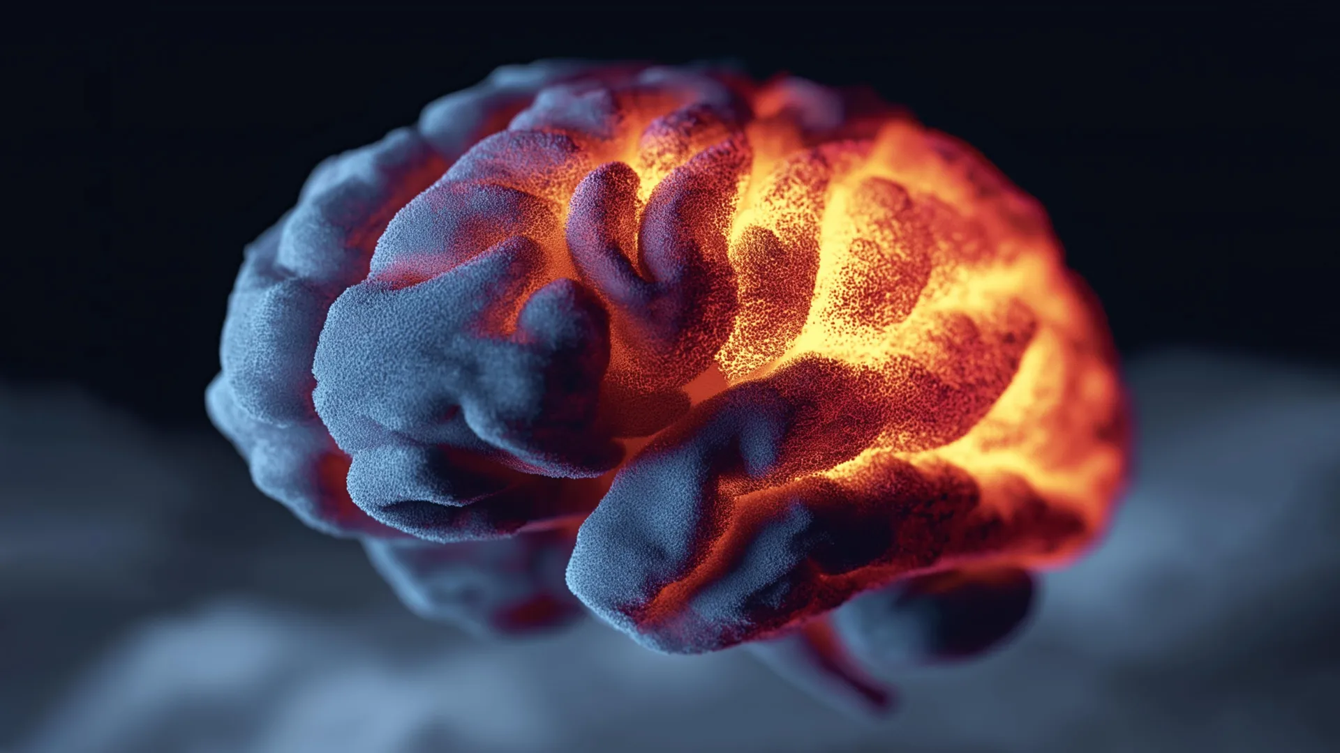
Unraveling the Mystery of Anti-NMDA Receptor Encephalitis: The Disease Behind "Brain on Fire"
While the gripping narrative of "Brain on Fire" brought anti-NMDA receptor encephalitis into the public consciousness, the disorder remains relatively rare, affecting an estimated 1 in 1 million people each year. Despite its rarity, the condition’s severe and often dramatic presentation, particularly in adults in their 20s and 30s, underscores the urgent need for improved diagnostics and treatments. Patients experience a rapid onset of severe neurological and psychiatric symptoms, often leading to misdiagnosis as a primary psychiatric illness, a delay that can have profound and lasting consequences.
The illness manifests when the body’s immune system, normally tasked with defending against foreign invaders, mistakenly turns against its own healthy tissues. In this specific disorder, the immune system targets NMDA (N-methyl-D-aspartate) receptors located in the brain. These receptors are not merely passive components; they are vital conduits for communication between neurons, playing an indispensable role in critical brain functions such as learning, memory formation, synaptic plasticity, and overall cognitive processing. The attack on these receptors is primarily driven by specific immune proteins known as anti-NMDA receptor autoantibodies.
The clinical presentation of anti-NMDA receptor encephalitis is notoriously varied and often terrifying for both patients and their families. Initial symptoms can be subtle, including headaches, fever, or mild personality changes, but they quickly escalate. Patients may experience profound and rapid cognitive decline, severe memory loss, and a range of dramatic psychiatric disturbances such as psychosis, paranoia, hallucinations, and agitated or catatonic states. Seizures are common, as are movement disorders (dyskinesias), speech disturbances, and autonomic instability, which can lead to life-threatening fluctuations in heart rate, blood pressure, and body temperature. In its most severe forms, the disease can progress to coma and, tragically, even death if left untreated or if treatments are ineffective. The rapid progression and the severity of symptoms highlight the critical need for early and accurate diagnosis, followed by swift and effective therapeutic intervention.
Pinpointing the Antibody Binding Sites: A New Therapeutic Avenue
The new OHSU study represents a significant methodological breakthrough, moving beyond a general understanding to pinpoint the precise molecular interactions that drive the disease. Scientists meticulously identified specific locations on a subunit of the NMDA receptor where these harmful autoantibodies attach. This level of precision is revolutionary because blocking these exact binding sites could potentially offer a highly targeted approach to slow or even reverse the progression of the disease, moving away from broad immunosuppression towards highly specific therapeutic interventions.
The lead author of the study, Junhoe Kim, Ph.D., a postdoctoral fellow at the OHSU Vollum Institute, spearheaded the intricate analysis. Dr. Kim’s research involved analyzing anti-NMDA receptor autoantibodies extracted from a specially engineered mouse model designed to mimic the human disease. Crucially, these findings were then rigorously compared with detailed, near-atomic resolution images of the same types of autoantibodies collected directly from human patients diagnosed with anti-NMDA receptor encephalitis.
The convergence of findings between the animal model and human subjects provided compelling validation for the research. The binding locations observed in the mouse model closely and consistently matched those identified in human patients. This cross-species validation is paramount in translational research, providing "really solid evidence," as emphasized by senior author Eric Gouaux, Ph.D., a senior scientist in the Vollum Institute and an investigator with the Howard Hughes Medical Institute. "We’re focused now on this area as literally a hot spot for the interaction that underpins at least one component of the disease," Dr. Gouaux stated, highlighting the critical importance of this newly identified region.
Dr. Kim further elucidated the advancement represented by their work, explaining that while earlier research had successfully narrowed down the general region where these antibodies might attach, his team’s study achieved unprecedented specificity. "From previous studies, people knew where the antibodies might bind," he said. "But we collected the entire native autoimmune antibody panel from a mouse model with the disease, and we elucidated where specifically they bind onto the receptor." This distinction between general localization and specific binding site identification is crucial, akin to knowing a particular city versus having the precise street address and building number.
Near-Atomic Imaging Reveals a Critical Hot Spot: The Power of Cryo-EM
Achieving such an extraordinary level of detail was made possible through the application of advanced near-atomic imaging techniques, specifically cryo-electron microscopy (Cryo-EM). The team utilized the state-of-the-art facilities at the Pacific Northwest Cryo-EM Center, strategically located on OHSU’s South Waterfront campus. This center is one of only three national facilities dedicated to this cutting-edge imaging technology, a testament to its importance and the specialized expertise required for its operation. It is a collaborative venture, jointly operated by OHSU and the Pacific Northwest National Laboratory, and significantly supported by the National Institutes of Health.
Cryo-EM allows researchers to visualize biological molecules, such as proteins and antibodies, in their near-native state and at incredibly high resolutions, sometimes down to individual atoms. Samples are rapidly frozen, preserving their structure, and then bombarded with electrons. The patterns of scattered electrons are then used to reconstruct a 3D image of the molecule, revealing its intricate architecture and, crucially, where other molecules might interact with it. This technology was indispensable in allowing the OHSU team to precisely map the antibody-receptor interaction.
Their meticulous analysis using Cryo-EM yielded a truly remarkable finding: nearly all of the pathogenic antibodies concentrated on a single, highly specific region, or domain, of the NMDA receptor. This concentration on a single domain is a "super exciting result," according to Dr. Gouaux, primarily because this particular part of the receptor happens to be the simplest to target therapeutically. The implications are profound, as a highly localized and accessible binding site simplifies the drug discovery process significantly, offering a clear molecular target for intervention.
Toward More Precise Treatments and Earlier Detection
The discovery of this "hot spot" has transformative implications for the future of anti-NMDA receptor encephalitis treatment. Co-author Gary Westbrook, M.D., a neurologist and senior scientist at the Vollum Institute, emphasized that this breakthrough could empower pharmaceutical companies to design novel drugs that specifically block these damaging antibody interactions.
Current treatments for anti-NMDA receptor encephalitis largely rely on broad immunosuppression. These therapies, which include corticosteroids, intravenous immunoglobulin (IVIg), and plasmapheresis, aim to dampen the overall immune response or remove circulating antibodies from the blood. While effective for many patients, these non-specific approaches come with significant limitations. They can leave patients vulnerable to infections due have widespread effects on the immune system, and they do not work for everyone. Furthermore, a substantial percentage of patients experience relapses, highlighting the need for more durable and targeted solutions.
"More specific approaches are definitely needed," Dr. Westbrook asserted. The OHSU findings pave the way for a new generation of precision medicines. Instead of broadly suppressing the immune system, future therapies could be designed to specifically interfere with the interaction between the autoantibodies and the NMDA receptor at the identified binding site. This could involve small molecule inhibitors that physically block the site, or even engineered antibodies (monoclonal antibodies) that act as "decoy receptors" or directly neutralize the pathogenic autoantibodies without broadly compromising immune function. Such targeted therapies promise fewer side effects, potentially higher efficacy, and a reduced risk of relapse, leading to better long-term outcomes and an improved quality of life for patients.
Beyond treatment, the research also holds immense promise for earlier and more accurate diagnosis. The ability to identify specific binding signatures of these autoantibodies at a molecular level could facilitate the development of a highly sensitive and specific blood test. Currently, diagnosing anti-NMDA receptor encephalitis can be challenging due to the diverse and often psychiatric nature of initial symptoms, leading to delays and misdiagnoses. An early blood test capable of detecting these specific antibody signs would allow clinicians to identify the disease much sooner, enabling patients to begin treatment earlier. This is critical, as early intervention is strongly associated with better neurological recovery and reduced morbidity. By preventing prolonged inflammation and damage to NMDA receptors, early treatment could significantly mitigate the devastating long-term effects of the disease.
Broader Impact and Collaborative Excellence
This research transcends the immediate context of anti-NMDA receptor encephalitis, offering a blueprint for understanding and treating other autoimmune neurological disorders. Many conditions, such as MOG antibody-associated disease (MOGAD) or neuromyelitis optica spectrum disorder (NMOSD), involve the immune system attacking specific brain or spinal cord components. The methodologies and insights gained from this study could potentially be applied to unravel the molecular mechanisms of these related conditions, accelerating the development of similar precision therapies across a spectrum of autoimmune encephalitides and myelopathies.
The success of this study is a testament to the power of collaborative, interdisciplinary scientific inquiry. The research team included Farzad Jalali-Yazdi, Ph.D., and Brian Jones, Ph.D., both from OHSU, alongside Drs. Kim, Gouaux, and Westbrook. This pooling of diverse expertise, from molecular biology and structural biology to clinical neurology, was essential in navigating the complexities of the research.
The study received significant financial backing from multiple prestigious organizations, including the National Research Foundation of Korea (award RS202400334731), the National Institute of Mental Health and the National Institute of Neurological Disorders and Stroke (both part of the National Institutes of Health, under award numbers F32MH115595, R01NS117371, and R01NS038631), the Howard Hughes Medical Institute, and Jennifer and Bernard LaCroute. Such robust support is vital for sustaining the high-cost, high-impact research that pushes the boundaries of medical science. It is important to note that the content presented is solely the responsibility of the authors and does not necessarily represent the official views of the NIH.
Furthermore, all animal research conducted at OHSU adheres to stringent ethical guidelines, undergoing rigorous review and approval by the university’s Institutional Animal Care and Use Committee (IACUC). The IACUC plays a crucial role in ensuring the welfare of animal subjects and the safety of research personnel, while also evaluating the scientific merit of all proposed animal studies to justify the use of live animals, ensuring that such research is both necessary and conducted with the highest ethical standards.
This landmark research from OHSU represents a pivotal moment in the fight against anti-NMDA receptor encephalitis. By precisely identifying the Achilles’ heel of the disease – the specific binding site of the pathogenic antibodies – scientists have opened new, highly promising avenues for developing targeted therapies and earlier diagnostic tools. This discovery offers not just scientific advancement, but genuine hope for a future where this devastating condition can be more effectively treated, and potentially even prevented, improving countless lives worldwide.