
 By admin
Dr. Martin Wessendorf, a distinguished neuroscientist and associate professor emeritus at the University of Minnesota, whose extensive research has focused on the intricate mechanisms by which the brainstem modulates neurons within the spinal cord, collaborated with Daniel Wang, a specialist in cognitive neuroscience and neurodegenerative diseases. Their combined expertise has allowed them to delve deeply into the evolutionary origins of the brain’s mysterious crossed connections, offering a compelling new hypothesis that reshapes our understanding of neuroanatomy.
In a groundbreaking study published in Frontiers in Neuroanatomy, Wessendorf and Wang meticulously examined several key neural circuits across a diverse range of four-limbed animals, or tetrapods. These circuits, universally exhibiting a cross-wired configuration where one side of the brain invariably connects with the opposite side of the body, form the bedrock of their investigation. Their hypothesis posits that these pervasive contralateral connections did not emerge as a direct, independent adaptation for efficiency or superior function, but rather developed as evolution strategically built newer, more complex brain circuits around an ancient, pre-existing visual system that was already inherently cross-wired. This suggests a profound instance of evolutionary repurposing, where an initial organizational principle dictated the subsequent development of vast swathes of the nervous system.
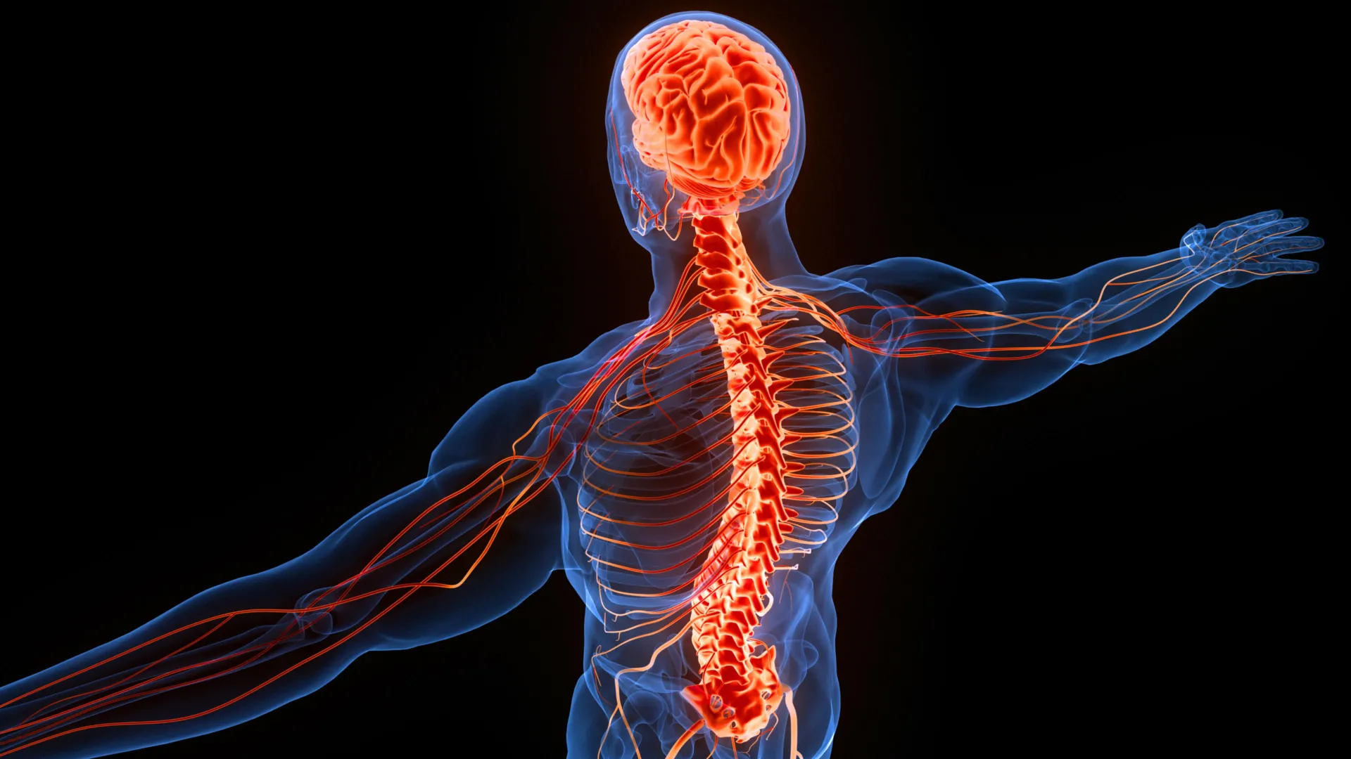
The vertebrate brain is a complex tapestry woven with numerous pathways that meticulously cross from one side of the central nervous system to the other. To truly grasp the enduring prevalence of these contralateral, or midline-crossing, connections, it is imperative to trace their development through the immense sweep of evolutionary time, examining how distinct sensory and movement circuits progressively emerged and integrated over hundreds of millions of years.
The Ancient Blueprint: How the Brain Processes Visual Information
One of the most powerful and enduring explanations for the brain’s crossed organization begins with the fundamental mechanics of vision. When light enters the eye and passes through its convex lens, the image projected onto the retina—the light-sensitive tissue at the back of the eye—is optically inverted both vertically and horizontally. Consequently, objects situated to our right are projected onto the left side of the retina, while objects on our left are projected onto the right. This initial optical inversion is a universal physical phenomenon, not a biological adaptation, but it sets the stage for the brain’s subsequent processing.
This inverted visual information then embarks on a critical journey along the optic nerve, leading to a pivotal structure known as the optic chiasm. Located ventrally in the diencephalon, this anatomical crossroads is where nerve fibers originating from the nasal (inner) halves of both retinas cross to the opposite side of the brain, while fibers from the temporal (outer) halves remain on the same side. The net effect of this precise organization is that visual information from the entire right visual field (perceived by the left half of each retina) is directed to the left side of the brain, and conversely, information from the entire left visual field (perceived by the right half of each retina) is directed to the right side of the brain. For most non-mammalian vertebrates, this crossing is often complete, meaning all visual information decussates. In mammals, partial decussation allows for binocular vision and depth perception.
A significant portion of these visual signals subsequently reaches an evolutionarily ancient brain structure called the optic tectum. In mammals, this structure is known as the superior colliculus, a prominent component of the midbrain. The optic tectum is not merely a relay station; it is a sophisticated center crucial for producing rapid, reflexive reactions to visual stimuli, such as swiftly orienting toward a sudden movement in our periphery. It acts as a primary sensorimotor interface, translating visual input into immediate motor responses.
Critically, each side of the optic tectum contains a topographical representation of the opposite visual field. This contralateral mapping is fundamental. Deeper within the optic tectum’s intricate layers, information pertaining to other physical sensations (somatosensory information) and auditory cues is also spatially organized into corresponding maps that align precisely with its visual representation. This multimodal integration, where various sensory inputs converge in a spatially consistent, contralateral manner, is a cornerstone of Wessendorf and Wang’s hypothesis. Because the optic tectum (or its homolog) is present in every vertebrate species, from the earliest fish to modern humans, it is considered one of the most ancient and conserved structures in the vertebrate nervous system. Its deep evolutionary roots and inherently contralateral organization may be particularly significant for understanding why so many other, evolutionarily newer neural pathways subsequently adopted a similar crossed architecture.
Sensory Signals: The Cross-Wired Paths to Perception
Wessendorf and Wang’s research further illuminates two major pathways responsible for relaying somatic sensations from the body to the brain, both of which demonstrate a characteristic crossing within the central nervous system (comprising the brain and spinal cord). These pathways appear to be widely conserved across tetrapods, underscoring their evolutionary importance.
The first, known as the dorsal column-medial lemniscal (DCML) pathway, is responsible for transmitting highly discriminative sensory information: gentle touch, vibration, and proprioception (the sense of the relative position of one’s own body parts and strength of effort used in movement). Signals originating from sensory receptors in the limbs and torso ascend ipsilaterally (on the same side) through the dorsal portion of the spinal cord in structures called the fasciculus gracilis and cuneatus. These fibers reach their first synapse in the brainstem, specifically in the nucleus gracilis and nucleus cuneatus within the medulla oblongata, just above the spinal cord. It is at this point that the pathway truly decussates: second-order neurons from these nuclei cross the midline as internal arcuate fibers, forming the medial lemniscus, which then ascends contralaterally to the thalamus, and subsequently to the primary somatosensory cortex.
While primitive fish generally lack a distinct dorsal column pathway, scientists have identified its presence and characteristic decussation in amphibians, reptiles, birds, and mammals. Its broad distribution across these major tetrapod groups strongly suggests that this precise arrangement emerged early in tetrapod evolution and has remained remarkably conserved, hinting at its adaptive significance for complex terrestrial locomotion and manipulation.
The second crucial pathway is the spinothalamic tract, also known as the anterolateral system. This pathway is responsible for transmitting sensations involving pain (nociception), temperature, and crude (non-discriminative) touch. Unlike the dorsal column pathway, the spinothalamic tract exhibits decussation much earlier in its journey. First-order neurons, originating from peripheral receptors, enter the spinal cord and synapse with second-order neurons in the dorsal horn. These second-order neurons then immediately cross to the opposite side of the spinal cord via the anterior white commissure, ascending contralaterally to the thalamus. From the thalamus, third-order neurons project to the somatosensory cortex.
This distinct crossed arrangement of the spinothalamic tract is found not only in birds, mammals, and reptiles but also occurs in some cartilaginous fish, such as sharks and rays. This presence in evolutionarily older fish lineages suggests that aspects of this organizational principle have even deeper evolutionary roots than the dorsal column pathway, potentially tracing back to the earliest gnathostomes (jawed vertebrates).
Motor Control: Orchestrating Movement Across the Midline
The pervasive crossed organization of the nervous system extends profoundly beyond sensation, deeply influencing how the brain orchestrates movement. Wessendorf and Wang identified three pivotal descending pathways involved in controlling skeletal muscles that consistently connect one side of the brain with the opposite side of the spinal cord, demonstrating a unified principle of contralateral motor control.
The first is the tectospinal pathway, a critical circuit that facilitates rapid, reflexive postural adjustments and orienting movements in response to sudden environmental cues. As previously noted, the optic tectum (superior colliculus in mammals) is a multimodal integration center. It processes not only visual information but also receives and integrates information about sounds and physical sensations, organizing these diverse signals into coherent spatial maps that align with its contralateral visual representation.
Consider a scenario: you are about to cross a busy street when, from the corner of your eye, you suddenly detect a car rapidly approaching. Without conscious thought, you instinctively turn your head and body toward the perceived threat. The tectospinal pathway is one of the primary neural circuits underlying this immediate, reflexive response. It originates in the deepest layers of the optic tectum, where multimodal sensory input converges, and its nerve fibers descend, crossing to the opposite side of the brainstem and spinal cord. This pathway then directly influences motor neurons and interneurons, coordinating the rapid turning of the head, eyes, and trunk. This evolutionarily ancient pathway is present in most tetrapods and even in certain cartilaginous fish, highlighting its fundamental role in survival.
Another significant motor circuit is the rubrospinal pathway, which is thought to play a crucial role in controlling limb movements, particularly those involving upper limb flexion and fine manipulation. This pathway originates in a distinctive midbrain structure called the red nucleus. From the red nucleus, its fibers promptly decussate (cross to the opposite side) in the ventral tegmental decussation and descend through the brainstem and spinal cord, influencing motor neurons. The rubrospinal pathway is especially prominent in most mammals, contributing to motor coordination and fine control, although it has also been identified in other tetrapods and some cartilaginous fish, suggesting its evolutionary lineage predates the full development of the corticospinal tract. In primates, it works in concert with the corticospinal tract, particularly for movements of the distal limbs.
The Evolution of Fine Motor Control in Humans
The third and perhaps most critical descending motor pathway for humans is the corticospinal tract, also known as the pyramidal tract. Its unique significance lies in its unparalleled role in mediating precise, voluntary movements, including the highly fractionated and delicate control of our fingers and hands, which is essential for tool use, writing, and intricate tasks.
The evolutionary history of the corticospinal tract is considerably more complex and recent than that of the other pathways. All vertebrates possess a telencephalon, the forward-most portion of the brain that accounts for a substantial proportion of the brain’s overall size in humans. However, only mammals possess a cerebral cortex that is organized into six distinct layers, a structure that forms the anatomical basis for much of our advanced cognitive abilities.
This six-layered neocortex supports an array of sophisticated functions, including the integration of sensory information with complex memories, the coordination of highly nuanced movements, and the processing of language. The corticospinal pathway originates from neurons in various cortical areas, primarily the primary motor cortex (M1), but also premotor, supplementary motor, and somatosensory cortices. Its fibers descend through the internal capsule, cerebral peduncles, and pons, eventually forming the pyramids of the medulla. It is at the medullary pyramids that the vast majority (approximately 85-90%) of these fibers famously decussate, forming the lateral corticospinal tract, which then descends contralaterally to innervate motor neurons in the spinal cord. The remaining uncrossed fibers form the anterior corticospinal tract, which typically decussates at the level of the spinal cord segments it innervates.
The function of the corticospinal pathway has evolved significantly across different animal groups. In most non-primate mammals, its primary influence is often indirect, acting mainly to modulate sensory processing or influencing spinal cord interneurons rather than directly innervating motor neurons. This allows the brain to regulate incoming sensations and influence general motor patterns. However, in primates, including humans, and certain other animals capable of highly precise movements (e.g., raccoons), the corticospinal pathway develops direct connections with alpha motor neurons in the spinal cord. These direct corticospinal connections are what make intricate, fractionated actions possible, enabling independent finger movements and the fine motor control needed for delicate tasks.
The researchers propose a particularly insightful hypothesis regarding the development of advanced fine motor abilities in humans. They suggest that instead of evolving an entirely new neural system for this purpose, humans may have developed these capabilities by adapting and repurposing an older, pre-existing neural circuit that was originally involved in controlling sensory input or more generalized motor patterns. In essence, evolution may have bestowed an existing, contralaterally organized pathway with an additional, more specialized purpose, rather than designing a de novo system. This exemplifies a recurring theme in evolution: modification of existing structures rather than wholesale creation.
A 500-Million-Year-Old Explanation for the Brain’s Crossed Wiring
When considered collectively, the intricate network of these neural pathways—visual, sensory, and motor—points toward a potentially surprising and remarkably deep evolutionary explanation for the brain’s consistently crossed organization. The researchers propose that the very earliest visual circuits, particularly those involving the optic chiasm and optic tectum, played a foundational role in establishing an initial, inherently crossed pattern. This fundamental organizational principle then served as a template, influencing how subsequent sensory and movement pathways were structured across opposite sides of the nervous system as they evolved, particularly within the lineage of tetrapods.
Given that every living vertebrate, from the most primitive fish to the most complex mammals, possesses an optic chiasm and an optic tectum organized around information from the opposite visual field, Wessendorf and Wang suspect that these structures, along with their contralateral organization, were already present in primitive jawless fish during the Cambrian Period, a geological epoch spanning approximately 538 to 486 million years ago. This era witnessed an explosion of life forms and the emergence of the earliest vertebrates.
If this hypothesis is correct, it would imply that the basic organizational principle underlying the brain’s profound crossed connections began developing more than half a billion years before the appearance of modern humans. This would make it one of the most ancient and enduring features of our nervous system. The hypothesis is not purely theoretical; it is potentially testable. Scientists could meticulously examine fossilized skulls from ancient jawless fish, such as those of the Ostracoderms or early Agnatha, for anatomical evidence. Such evidence might include the size and positioning of optic nerve foramina or subtle impressions within the braincase that could help establish whether these early animals possessed structures analogous to an optic chiasm and an optic tectum, and how they might have been organized.
Evolutionary Inheritance Rather Than Direct Advantage?
The broader implications of this hypothesis challenge a common assumption in evolutionary biology: that every unusual or prominent biological feature must have evolved because it conferred a direct and immediate survival benefit. While adaptation is undeniably a powerful force, evolution frequently operates by modifying structures and pathways that already exist, rather than consistently developing entirely new systems from scratch.
Once the ancient visual pathways established their characteristic crossed organization—a necessity driven by the physics of light and the initial optical inversion on the retina—that fundamental arrangement may have exerted a powerful and lasting influence on how subsequent, more complex additional sensory and motor circuits developed. In particular, the multimodal integration and contralateral mapping within the optic tectum likely provided a robust structural and functional foundation that later pathways found advantageous or simply "easier" to follow and build upon.
Under this compelling hypothesis, the brain’s crossed connections did not necessarily arise because controlling the opposite side of the body was inherently a more effective or efficient strategy for all functions. Instead, they may largely reflect the enduring legacy of an ancient organizational principle—an "evolutionary inheritance"—that was repeatedly reused, adapted, and elaborated upon throughout vertebrate history. The result is one of the most profound and enduring examples of how the nervous system faithfully preserves its deep evolutionary history. The strange yet fundamental relationship between the brain’s two hemispheres and the opposite sides of the body may ultimately trace its origins back to the very visual systems of animals that swam the primeval oceans more than 500 million years ago, a testament to the conservatism and ingenuity of natural selection.
