
 By admin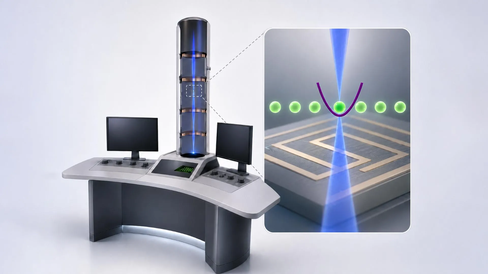
For decades, electron microscopy has stood as a cornerstone of scientific discovery, pushing the boundaries of what we can observe at the atomic and molecular scales. From unveiling the intricate architecture of viruses to mapping the precise arrangement of atoms in advanced materials, these powerful instruments have revolutionized fields ranging from biology and medicine to materials science and nanotechnology. Unlike optical microscopes that use photons of light, electron microscopes employ a beam of electrons, which, due to their much shorter de Broglie wavelength, can resolve details far beyond the diffraction limit of visible light. However, despite their extraordinary capabilities, the fundamental principle behind image formation in most conventional electron microscopes—the simple counting of scattered electrons—might be inherently limiting their potential, discarding a wealth of quantum information each electron carries.
This is the premise of a groundbreaking new approach being developed by a consortium of Austrian researchers. Their vision is to transcend the classical limitations of electron microscopy by integrating it with the cutting-edge capabilities of quantum computing. By doing so, they aim to tap into the hitherto unused quantum information carried by each electron, promising to yield clearer images from fewer electrons and unlock new insights into the most delicate of samples. The collaborative effort, involving experts from TU Wien, the University of Vienna, JKU Linz, and the University of Innsbruck, represents a bold step towards a new era of quantum-enhanced imaging, with a prototype quantum computer electron microscope already under construction at TU Wien.
The Unmet Challenge: The Electron Budget and Sample Damage
Conventional electron microscopes operate on the principle of detecting electrons that have interacted with a sample. In a standard transmission electron microscope (TEM), electrons pass through a thin sample, and the variations in their scattering (due to differences in atomic number, density, and thickness) are used to form an image. Scanning electron microscopes (SEM) and scanning transmission electron microscopes (STEM) raster a focused electron beam across the sample surface or through it, respectively, detecting various signals (secondary electrons, backscattered electrons, transmitted electrons) to build an image pixel by pixel. The resolution achieved by these instruments is truly remarkable, with modern aberration-corrected STEMs capable of resolving individual atoms, pushing into the sub-angstrom regime.
"Today, we can image tiny details on the atomic scale," affirms Philipp Haslinger from the Institute of Atomic and Subatomic Physics at TU Wien. This extraordinary resolution has, for instance, been pivotal in structural biology, where techniques like cryo-electron microscopy (cryo-EM) have enabled the visualization of complex biological macromolecules, earning its developers a Nobel Prize. By flash-freezing samples, cryo-EM mitigates some of the damage, allowing researchers to average thousands or even millions of images of identical molecules to reconstruct a high-resolution 3D structure.
However, a fundamental dilemma persists: the very electrons used to image a sample can also damage it. Electrons are highly energetic particles, and their interaction with matter can lead to ionization, bond breaking, mass loss, and structural rearrangements within the sample. This phenomenon, known as "radiation damage" or "beam damage," imposes a severe "electron budget" – a limit on the total number of electrons a sample can withstand before its structure is irrevocably altered.
"However, this requires a large number of electrons. And not every sample can be exposed to so many electrons without being damaged. This is often a problem, particularly when imaging biological samples such as individual proteins," Haslinger emphasizes. Biological materials, composed primarily of light elements and complex organic molecules, are notoriously sensitive to electron beam radiation. Imaging a protein in its native, functional state often means navigating a tightrope between acquiring enough signal for a clear image and preserving the sample’s integrity. For many biological systems, especially those exhibiting conformational dynamics or intrinsic disorder, the electron budget is simply too restrictive, leading to blurred features or artifacts that obscure critical biological information. Even with cryo-EM, the damage is merely slowed, not eliminated, necessitating extensive averaging and computational reconstruction, which can itself obscure dynamic processes or unique structural features of individual molecules.
The challenge, therefore, is not merely to achieve higher resolution, but to learn more from each electron – to extract maximum information while minimizing the total electron dose. This would allow researchers to produce clearer images with fewer electrons, thereby extending the lifetime of delicate samples under the beam and revealing details currently lost to radiation damage or statistical noise.
Beyond Counting: Harnessing Quantum Entanglement
In a standard electron microscope, the electron is treated primarily as a classical particle, and its interaction with the sample is registered as a count at a detector. The quantum information it carries – its intrinsic properties and the subtle quantum correlations it develops through interaction with the sample – largely goes unnoticed and unused. The Austrian team’s revolutionary idea is to tap into this hidden quantum realm.
Their approach involves linking the electron microscope to a quantum computer built around trapped ions. This audacious fusion aims to transform the electron from a simple probe into a quantum messenger, whose subtle quantum state, altered by the sample, can be read and processed with unprecedented sensitivity.
"Our idea is to combine the electrons with a quantum computer. We let them interact with ions that are held in place along the path of the electron beam," explains Elias Pescoller, first author of the publication and a doctoral student at the Institute for Theoretical Physics and the Institute of Atomic and Subatomic Physics at TU Wien. The critical step here is the creation of quantum entanglement. Entanglement is a unique quantum phenomenon where two or more particles become linked in such a way that the quantum state of each particle cannot be described independently of the others, even when separated by vast distances. A measurement on one instantaneously influences the other.
In this setup, an electron that has just interacted with the sample then encounters a trapped ion. This interaction is engineered to create quantum entanglement between the electron and the ion. "This can, for example, create quantum entanglement between the electron and the quantum computer. The electron and the ion then share a joint quantum state," Pescoller elaborates. Essentially, information about the electron’s interaction with the sample – information encoded in its quantum state – is transferred to and stored within the quantum state of the ion.
Turning Weak Signals into Useful Information through Quantum Processing
The power of this system lies in its ability to accumulate and process quantum information over time. After one electron interacts with a trapped ion and entangles with it, another electron can follow suit, also becoming entangled with the quantum computer. By carefully orchestrating a sequence of quantum operations, the system can combine information from multiple electrons in a way that is fundamentally impossible with classical methods.
"If we perform very specific quantum-computing operations each time, we can optimally combine the information from several electrons so that we obtain a signal of maximum strength even though we use only a relatively small number of electrons," says Dennis Rätzel from the Institute of Atomic and Subatomic Physics at TU Wien. This is where the quantum computer truly shines. Classical signal processing relies on averaging noisy signals, where the signal-to-noise ratio improves with the square root of the number of measurements. However, this method is fundamentally limited by statistical fluctuations, often referred to as "shot noise," which becomes dominant at low electron doses.
Quantum computers, by contrast, can exploit quantum correlations to extract information that would otherwise be buried in noise. The algorithms necessary for this sophisticated processing were developed in collaboration with Johannes Kofler’s team at JKU Linz. These algorithms are designed to decode the entangled states, piecing together a coherent picture from faint, quantum-encoded signals that would appear as mere random fluctuations to a classical detector.
"The electrons themselves are used to image small objects, just as in any other electron microscope. But by processing the quantum information carried by these electrons in a quantum computer, we can extract significantly more information from the process," explains Iva Březinová from the Institute for Theoretical Physics at TU Wien. "What would previously have been indistinguishable from random noise can thus become a clear signal." This capability is transformative. It means that subtle interactions between electrons and the sample, which are crucial for understanding fine structural details or weak atomic potentials, can be amplified and rendered visible.
Elias Pescoller succinctly summarizes the profound implication: "Quantum physics allows us to overcome the statistical limits that constrain conventional electron microscopes." This ability to bypass classical statistical limitations opens up an entirely new regime for electron microscopy, where the trade-off between resolution, signal strength, and sample damage can be fundamentally re-evaluated.
From Mathematical Proof to a Working Quantum Microscope
The journey from a theoretical concept to a functional instrument is arduous, but the Austrian team has already made significant strides. Their initial work has provided the mathematical proofs demonstrating the profound advantages this quantum-enhanced method should offer. The next, and arguably most challenging, phase is the experimental realization of this vision.
At TU Wien’s University Service Center for Transmission Electron Microscopy (USTEM), researchers are now actively preparing for the ambitious task of integrating an ion-based quantum computer directly into an electron microscope. The quantum computer itself, a marvel of quantum engineering, was developed by Philipp Schindler’s team at the University of Innsbruck, renowned for their pioneering work in trapped-ion quantum systems. This integration involves overcoming formidable technical hurdles, including maintaining ultra-high vacuum conditions for both the electron microscope and the trapped ions, precise alignment of the electron beam with the ion trap, and ensuring the coherence of quantum states in a complex experimental environment.
If this pioneering system performs as anticipated, it promises to usher in a paradigm shift in electron microscopy. Researchers would gain the unprecedented ability to extract substantially more information from each electron, thereby enabling the visualization of structures and processes that are currently inaccessible due to radiation damage or insufficient signal. This would be particularly revolutionary for fields like structural biology, allowing scientists to image delicate proteins, viruses, and cellular components at near-native states with minimal perturbation, potentially even capturing dynamic conformational changes in individual molecules. In materials science, it could unlock the study of highly radiation-sensitive materials, advanced catalysts, or the subtle quantum phenomena within novel electronic materials.
The collaborative nature of this project, bringing together disparate fields of expertise, is key to its potential success. "It is really exciting that, within the quantA Cluster of Excellence, we can combine the expertise in quantum information, quantum computing and electron microscopy available at the different universities in Austria. This allows us to launch a unique project," says Thomas Juffmann from the University of Vienna, highlighting the synergy between theoretical physicists, experimentalists, quantum computing specialists, and microscopy experts. This interdisciplinary approach, fostered by significant funding from the Austrian Science Fund (FWF) through the Cluster of Excellence quantA and the prestigious Gordon and Betty Moore Foundation, underscores the global recognition of this project’s transformative potential.
The construction of this quantum computer electron microscope at TU Wien is not just an engineering feat; it represents a bold exploration into the quantum limits of imaging. Should it prove successful, this technology will not merely enhance existing capabilities but will open entirely new avenues of scientific inquiry, allowing us to peer deeper into the quantum fabric of matter with unparalleled clarity. The future of microscopy, it seems, is undeniably quantum.
