
 By admin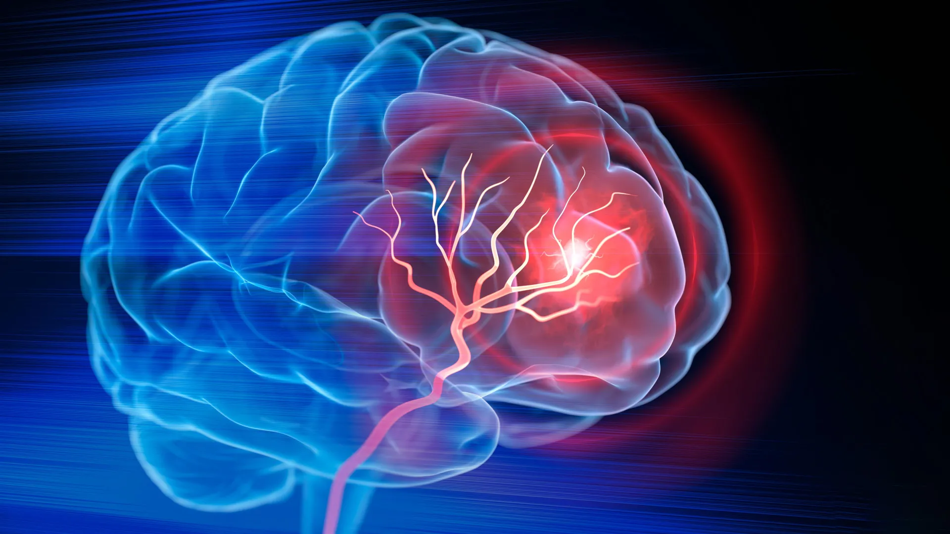
This groundbreaking research, recently published in the esteemed journal Cell Biomaterials, marks a significant step forward in addressing one of the most devastating neurological conditions globally. The innovative treatment demonstrated a remarkable capacity to orchestrate the body’s intrinsic repair mechanisms, actively recruiting the immune system, fostering the formation of vital new blood vessels, supporting profound changes in neural tissue architecture, and ultimately leading to measurable improvements in motor function in the animal models tested. These findings offer a beacon of hope for millions of individuals worldwide grappling with the aftermath of stroke, a condition that frequently leaves patients with profound and often permanent disabilities.
The Enduring Challenge of Stroke and Brain Repair
Stroke remains a leading cause of long-term disability and mortality across the globe. Each year, an estimated 15 million people suffer a stroke, with ischemic strokes accounting for approximately 87% of all cases. These occur when a blood clot obstructs blood flow to a critical region of the brain, depriving neurons of oxygen and nutrients. The consequences are immediate and often catastrophic, leading to rapid neuronal death in the core of the affected area and progressive damage in the surrounding "ischemic penumbra."
Current emergency treatments, such as clot-dissolving drugs (thrombolytics) and mechanical thrombectomy (physical clot removal), are vital for restoring circulation and salvaging brain tissue that is still viable. Administered within a narrow therapeutic window, these interventions can dramatically reduce the extent of initial brain damage. However, once brain tissue has succumbed to ischemia and died, restoring blood flow alone cannot regenerate what has already been lost. The brain, unlike many other organs, possesses a limited capacity for self-repair, particularly when faced with substantial tissue loss.
Severe ischemic strokes can devastate large areas of the brain, leaving behind a persistent cavity or lesion where healthy neural tissue once thrived. This void not only represents a physical absence of functional tissue but also disrupts complex neural networks, leading to a cascade of neurological deficits. Post-stroke recovery largely hinges on intensive rehabilitation therapies, which aim to help surviving brain circuits adapt and reorganize – a process known as plasticity – to compensate for lost function. While rehabilitation is indispensable and can yield significant functional improvements, it does not directly rebuild or regenerate the damaged brain region itself. The structural void often remains, posing a persistent challenge to complete recovery.
"Once brain tissue has been lost, simply restoring blood flow is no longer enough to achieve meaningful regeneration," stated Tatiana Segura, the Robert Plonsey Distinguished Professor of Biomedical Engineering at Duke University and senior author of the study. Her words underscore the critical unmet need in stroke treatment. "Our overarching goal is to engineer the injured space itself, transforming it into an environment where immune, vascular, and neural repair processes can begin to work in concert, fostering true regeneration rather than just adaptation." This paradigm shift – from simply preventing further damage to actively facilitating tissue regrowth – is at the heart of the Duke team’s innovative approach.
Building a Dynamic Scaffold for Brain Regeneration
Recognizing the multifaceted nature of brain repair, Professor Segura and her team embarked on a mission to create an internal environment within the stroke cavity capable of simultaneously supporting several crucial types of repair. Their solution involves a sophisticated biomaterial called MAPS, or microporous annealed particle scaffolds. These scaffolds are not monolithic structures but rather consist of individual hydrogel microparticles that spontaneously assemble into a highly porous, interconnected network upon injection.
Hydrogels are biocompatible, water-rich polymeric materials that can mimic the soft tissue environment of the brain. The brilliance of MAPS lies in its unique architecture: the open, interconnected spaces within the scaffold provide an intricate three-dimensional framework that cells can readily enter, colonize, and utilize as a guide for rebuilding neural tissue. This porous design is crucial, as it allows for cell infiltration, nutrient exchange, and the establishment of new cellular connections, all of which are essential for tissue regeneration.
Building on earlier successes with their biomaterial platform, the researchers sought to push the boundaries further by actively engaging the body’s own immune system to guide and strengthen the repair process. For this critical component of their strategy, they focused on astrocytes. These star-shaped glial cells are the most abundant cell type in the brain, playing indispensable roles in normal brain function, including supporting neuronal metabolism, regulating synaptic transmission, and maintaining the blood-brain barrier. Crucially, astrocytes are also among the first cells to respond robustly when the brain is injured, undergoing reactive astrogliosis, which can have both beneficial and detrimental effects.
A key mechanism through which astrocytes communicate with surrounding cells, both in health and disease, is by releasing extracellular vesicles (EVs). These extremely small, lipid-bilayer-enclosed packages act as sophisticated intercellular messengers, carrying a diverse cargo of proteins, lipids, messenger RNA, and microRNAs. EVs can influence the behavior of recipient cells by transferring their contents, making them powerful mediators of biological responses and attractive candidates for therapeutic delivery.
Precision Delivery: Keeping Repair Signals Localized
A significant challenge in regenerative medicine, particularly within the central nervous system, is ensuring that therapeutic signals reach their intended targets and remain localized long enough to exert their desired effects. Systemic administration of signaling molecules often leads to rapid degradation, off-target effects, and insufficient concentrations at the site of injury, especially given the formidable blood-brain barrier.
To overcome this, the Duke researchers adopted an ingenious strategy. They first collected EVs from lab-grown astrocytes, creating a rich source of these natural communication packages. They then meticulously tested these EVs in combination with different signaling molecules known for their potential to attract immune cells, encourage blood vessel repair, and improve neural function. However, instead of simply injecting the EVs directly into the damaged area, which might lead to their rapid diffusion and clearance, the scientists chemically attached them to the surfaces of the hydrogel microparticles within the MAP scaffold.
This elegant modification was pivotal. By anchoring the EVs to the scaffold, the therapeutic signals were kept concentrated within the lesion cavity, creating a high local density of pro-regenerative cues. This localized delivery significantly enhanced the probability that incoming cells would encounter these critical signals, maximizing their therapeutic impact. "We are not simply placing an inert material into the brain and hoping for the best," Professor Segura emphasized. "Instead, we are actively engineering a local environment that can precisely coordinate and amplify several crucial parts of the repair response, essentially creating a regenerative niche."
Among the various signaling combinations tested, one particular duo – Interleukin-4 (IL-4) and Complement component 1q (C1q) – demonstrated exceptional efficacy. This combination proved particularly potent at drawing potentially helpful immune cells into the injured region. These recruited cells included macrophages, known for their phagocytic and immunomodulatory roles, as well as a surprisingly persistent population of neutrophils.
An Unexpected Hero: The Role of Neutrophils in Repair
The discovery of a beneficial role for neutrophils in stroke recovery is one of the study’s most intriguing and potentially paradigm-shifting findings. Neutrophils are typically characterized as the frontline soldiers of the innate immune system, rapidly migrating to sites of acute inflammation and infection. In the context of ischemic stroke, neutrophils have historically been primarily linked to the early stages of injury, where their excessive activation and release of proteolytic enzymes and reactive oxygen species contribute significantly to inflammation, secondary tissue damage, and the exacerbation of brain injury. Their presence has often been considered detrimental.
However, the new results from the Duke team suggest a far more nuanced and complex role for these cells. At a later point after injury, and when meticulously guided by the right biomaterial environment and specific signaling molecules, neutrophils may, in fact, pivot from being agents of destruction to critical supporters of tissue repair.
To validate their hypothesis, researchers conducted experiments where they selectively reduced the population of these newly recruited, neutrophil-rich immune cells within the scaffold-treated stroke cavity. The outcome was stark: when neutrophils were diminished, there was a substantial decline in new blood vessel formation, and the biomaterial scaffold underwent significantly less remodeling and integration into the host tissue. This compelling result strongly indicated that these immune cells, previously viewed as largely harmful in stroke, were playing an indispensable and positive role in the healing response.
"This result fundamentally changes how we think about neutrophils after stroke," said Shangjing Xin, the lead scientist of the study and a postdoctoral fellow in the Segura Laboratory. His statement underscores the significance of the finding. "Their role appears to be highly context-dependent, influenced by when they arrive, where they are located within the injured brain, and critically, the specific signals they receive from their surroundings. Our study demonstrates a potential engineering strategy to precisely recruit and retain these cells at the right time and place, essentially reprogramming their function from detrimental to beneficial." This re-evaluation of neutrophil function opens new avenues for immunomodulatory therapies in stroke.
Restoring Function: New Blood Vessels and Neural Growth
The biological transformations orchestrated by the injectable biomaterial translated directly into tangible functional improvements. As immune cells infiltrated the treated area, a robust network of new blood vessels began to form throughout the stroke cavity. This process, known as angiogenesis, is critical for supplying oxygen and nutrients to the regenerating tissue, facilitating cellular survival and integration.
Furthermore, researchers observed a significant increase in the density of axonal fibers – the long, slender projections of neurons that transmit electrical signals – both within and surrounding the injured region. This axonal sprouting and potential regeneration are crucial indicators of neural plasticity and the brain’s attempt to re-establish disrupted communication pathways. The emergence of these new connections is a prerequisite for functional recovery.
These profound biological changes were accompanied by remarkable improvements in motor function in the animal models. Mice treated with the optimized biomaterial scaffold performed significantly better on a standardized grid-walking test, a behavioral assessment designed to precisely measure fine motor coordination and identify mistakes in forelimb placement. By eight weeks post-treatment, their performance on this challenging task could not be statistically distinguished from that of healthy, uninjured control mice – an exceptional outcome in preclinical stroke research. Crucially, this impressive functional improvement was not transient; it persisted for the remainder of the study, indicating a sustained and stable recovery.
The Indispensable Role of the Scaffold
To rigorously ascertain the necessity of the biomaterial scaffold, the researchers conducted a critical comparative experiment. They tested whether the extracellular vesicles (EVs) alone, administered without the MAP scaffold, could produce similar therapeutic effects. The results were unequivocal: EVs administered in the absence of the MAP scaffold failed to elicit comparable blood vessel repair or neural tissue remodeling.
This finding strongly suggests that the biomaterial was doing far more than simply acting as a passive vehicle for transporting therapeutic signals into the brain. Its unique porous architecture, combined with its ability to physically concentrate and retain the EV-borne signals within the damaged region, appeared to be absolutely critical to initiating and sustaining the complex repair response. The scaffold effectively created a localized, bio-active niche that fostered cellular infiltration, communication, and the intricate processes required for tissue regeneration. Without this engineered microenvironment, the therapeutic potential of the EVs was significantly diminished, highlighting the synergistic power of the combined approach.
A Glimpse into the Future: Translational Pathways and Next Steps
Despite the highly promising and exciting results, it is important to acknowledge that this approach remains in its preclinical stages. So far, researchers have rigorously tested the treatment in well-established mouse models of ischemic stroke, where the material was injected directly into the damaged area of the brain. While animal models provide invaluable insights, the journey from laboratory discovery to human clinical application is long and complex.
Further extensive research will be necessary to thoroughly assess the safety profile of the biomaterial and its components, to precisely understand the intricate mechanisms by which different immune-cell populations influence recovery, and to determine the efficacy and safety of the treatment in larger animal models that more closely resemble the human brain in terms of size and complexity. These steps are crucial for addressing potential challenges such as immune responses to the biomaterial, long-term stability, and the exact dosage and timing for optimal therapeutic effect.
Currently, the researchers obtain the extracellular vesicles from primary rat astrocytes. While effective for preclinical studies, this source presents limitations for clinical translation, including issues of scalability, reproducibility, and potential immunogenicity in human patients. As a critical next step, Professor Segura’s laboratory is actively investigating the use of EVs produced by human induced pluripotent stem cell (hiPSC)-derived astrocytes. Such cells offer a highly scalable, ethically sound, and clinically relevant source of EVs. Furthermore, using hiPSC-derived astrocytes could provide researchers with an unprecedented level of control over the specific signals contained inside the EVs, allowing for the engineering of even more potent and targeted therapeutic cargo.
Professor Segura eloquently encapsulates the philosophy behind their work, drawing an analogy to ecological restoration: "You do not restore a damaged ecosystem simply by containing the initial damage; you have to actively create the conditions that allow life to return and flourish. That is precisely how we conceptualize the stroke cavity. The material is not intended to reproduce the brain itself, which is an impossible feat. Instead, its purpose is to create an enabling environment – a regenerative niche – where the body’s own cells can enter, effectively communicate with each other, and actively participate in rebuilding functional, vascularized tissue." This holistic vision for regenerative medicine after stroke offers a powerful and inspiring direction for future therapeutic strategies.
