
 By admin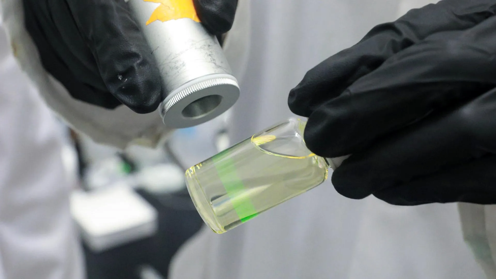
At its core, the technology relies on minuscule particles that, upon encountering a target chemical, emit a distinctive light signal. These nanoprobes possess the remarkable ability to absorb low-energy photons and transform that energy into higher-energy photons, generating a bright optical signal readily measurable by researchers. This "upconversion" phenomenon is a critical departure from conventional methods, offering profound advantages in detection.
The practical ramifications of this unusual capability are vast and varied. Pharmaceutical manufacturers, for instance, could deploy these particles to swiftly and accurately identify unwanted impurities or subtle structural variations in drug compounds—a critical step in ensuring efficacy and patient safety. Similarly, environmental scientists could utilize them to monitor for minute quantities of chemical pollutants in complex matrices like groundwater, providing an early warning system for contamination that is currently difficult and expensive to achieve. Beyond these immediate applications, the technology holds promise for biomedical diagnostics, industrial process control, and even advanced materials research.
Turning Infrared Light Into a Bright Green Signal
For decades, the field of optical sensing has predominantly relied on organic molecules known as fluorophores. These compounds absorb light and re-emit it at a different, usually lower, energy, producing vibrant, colorful emissions. However, as Professor Kai Huang, senior author on the paper describing the new particles published in the prestigious Journal of the American Chemical Society, explains, "Organic molecules called fluorophores have been used for decades to absorb light and convert it into colorful emissions, but the process only works in one direction."
Professor Huang elaborates on this fundamental limitation: "With fluorophores, the excitation frequency has to be higher than the emission frequency, which means that they convert high-energy photons into low-energy photons." This "downconversion" process, while widely used, comes with inherent drawbacks. High-energy excitation light (e.g., UV or visible blue light) can cause photobleaching, damaging the fluorophore over time, and often leads to significant background noise from the sample itself, known as autofluorescence. This autofluorescence can mask the faint signal from the target molecule, severely limiting detection sensitivity, especially in biological or environmental samples.
"What makes our dye-sensitized nanoparticles special is that they are capable of upconversion, meaning that they can absorb light in the form of low-energy photons and emit higher-energy ones," Professor Huang states. This inverse energy conversion is the cornerstone of their breakthrough. Instead of starting with high-energy light and ending with lower-energy light, these nanoparticles reverse the process. "For example, you could excite them with near-infrared light, which can easily be produced with low-cost lasers, and they would glow bright green in response."
This upconversion capability bestows several significant advantages. Near-infrared (NIR) light, being lower in energy, causes less photobleaching and has superior penetration depth into complex samples, making it ideal for applications in biological tissues or turbid environmental water samples. More critically, because the light used to activate them (NIR) has a vastly different frequency from the light they emit (green), researchers can effortlessly separate the desired signal from any background light or autofluorescence generated by the sample itself. This spectral separation is a game-changer for high-sensitivity detection.
Professor Huang likens the profound impact of this zero-autofluorescence background to a celestial observation. "It’s like the difference between stargazing at night versus the daytime," he says. He further clarifies, "The stars shine the same brightness all the time, but during the day the sun is so powerful that it overwhelms them. Shifting the excitation frequency lower produces zero-autofluorescence background in the samples you are analyzing, while the luminescent nanoprobes keep shining; it is like turning off the sun, so you can see the stars better." This eloquent analogy perfectly encapsulates how the upconversion mechanism effectively eliminates the "sun" of background noise, allowing even the faintest "stars" of target signals to shine brightly and clearly.
The Challenge of Making Nanoparticles Brighter
The innovative upconversion process within these nanoparticles is orchestrated by ions of ytterbium (Yb) and erbium (Er), two elements belonging to the lanthanide family of rare-earth metals. Lanthanides are renowned for their unique and highly efficient optical properties, making them ideal candidates for light-emitting applications. In this system, ytterbium ions act as a sensitizer or "relay," efficiently absorbing the incoming energy, while erbium ions serve as the "emitter," performing the actual upconversion and emitting the higher-energy light.
Earlier iterations of these sensing nanoparticles typically featured a relatively simple, flat hexagonal structure. In these designs, the ytterbium and erbium ions were homogeneously distributed throughout a host material, often composed of sodium, yttrium, and fluorine. The researchers compared this arrangement to "chocolate chips embedded in a cookie," with organic dye molecules resembling "icing" covering the outside surface. These organic dyes play a crucial role in "sensitizing" the nanoparticles, enhancing their initial absorption of the low-energy infrared light.
When near-infrared light irradiates these dye-sensitized nanoparticles, the organic dyes first capture the incoming energy with high efficiency. This captured energy is then transferred to the ytterbium ions, which act as energy relays, accumulating and channeling it. Subsequently, this accumulated energy is passed from the ytterbium ions to the erbium ions. It is within the erbium ions that the critical upconversion step occurs, leading to the emission of higher-energy photons, which manifest as bright green light.
However, the quest for ever-brighter and more efficient upconversion systems has historically encountered a significant hurdle. As Jiaze Wu, a PhD student in Professor Huang’s lab and the lead author on the new paper, explains, "But there’s a problem: if you pack the ytterbium atoms in too densely, they start to absorb not only the energy coming in, but also the energy coming out." This phenomenon, known as "back-energy transfer" or concentration quenching, severely limits the overall brightness and efficiency of the nanoparticle.
Wu further clarifies the detrimental impact: "This is called back-energy transfer: it means that the energy that would have been emitted by the erbium ions as green light instead gets bounced back to the ytterbium relay and never reaches the surface." In essence, the densely packed ytterbium ions, while excellent at collecting energy, also become proficient at re-absorbing the very light that the erbium ions are trying to emit, effectively trapping the signal within the nanoparticle and preventing it from being detected. Overcoming this fundamental limitation was paramount to achieving high-performance sensing.
A Layered Design Creates a One-Way Energy Path
To circumvent the problem of back-energy transfer and unlock the full potential of upconversion, Wu, Professor Huang, and their multidisciplinary team embarked on a comprehensive redesign, targeting both the chemical composition and the physical structure of the nanoparticles. Their innovative approach yielded a multi-layered architecture that fundamentally alters energy flow within the particle.
First, they moved beyond the traditional host matrix of sodium, yttrium, and fluorine. The researchers engineered a new matrix utilizing lithium, lutetium, and fluorine. This new composition provides a more optimized environment for the lanthanide ions, potentially reducing lattice defects and improving energy transfer efficiency, while also allowing for higher doping concentrations without detrimental effects.
Concurrently, they transformed the particles’ morphology from flat, two-dimensional hexagons into more intricate, three-dimensional diamond-shaped structures. The most significant structural innovation, however, lies in the internal architecture: each particle is composed of several distinct, concentric regions—a dense core encapsulated by an inner shell, which is then further enveloped by an outer shell.
This sophisticated layering allows for precise control over the distribution of the sensitizer ions. "We were able to create nice gradient: the concentration of embedded ytterbium ions gets denser as you go through each layer, with the core being the most dense," says Wu. This strategic gradient is the key to preventing back-energy transfer. By concentrating the ytterbium ions more densely towards the core and progressively less so towards the outer layers, the energy transfer pathway is effectively directed.
"This arrangement enabled us to pack in much more ytterbium," Wu explains, "In our particles, the light energy coming in flows almost entirely in one direction, inward toward the erbium ions." This ingenious core-shell-shell design, with its carefully controlled ytterbium gradient, ensures that energy is efficiently captured and channeled inwards to the erbium emitters, and once upconverted, the emitted light can escape without being re-absorbed by the ytterbium sensitizers. It creates a highly efficient, one-way "energy tunnel."
Crucially, this complex design was not the result of arduous trial and error alone. The team heavily leveraged advanced computational modeling techniques to accelerate their discovery process. Researchers simulated dozens of potential chemical formulations and particle shapes, predicting their energy transfer dynamics and optical properties, before committing to the laboratory synthesis of only the most promising versions.
Undergraduate student Weixiang Ben, who spearheaded the computational efforts, highlights the depth of their simulations: "We used Monte Carlo simulations and density functional theory to simulate how the energy would interact between different parts of the nanoparticle, right down to the atomic or even subatomic level." Monte Carlo simulations, which use random sampling to model complex systems, helped predict energy migration pathways, while Density Functional Theory, a quantum mechanical modeling method, provided insights into electronic structure and energy levels. "That’s how we showed that this core-shell-shell structure could actually function as a one-directional energy tunnel for incoming light," Ben affirms. This synergistic approach of computational design and experimental validation drastically reduced development time and optimized performance.
Nanoparticles Up to 150 Times Brighter
The culmination of this meticulous design and computational optimization is a new class of upconversion nanoparticles that produce an immensely stronger signal than any previous iteration. The performance metrics reported by the University of Toronto team are truly remarkable.
According to Jiaze Wu, the emitted light from their novel nanoparticles is approximately 150 times brighter than that of upconversion nanoparticles that have not been dye-sensitized. This highlights the critical role of the organic dyes in boosting the initial absorption of infrared light, significantly enhancing overall efficiency. Furthermore, under identical excitation conditions, these newly developed nanoparticles are about 50 times brighter than even some of the most highly optimized conventional upconversion structures previously documented in scientific literature. This comparison underscores the transformative impact of their layered architecture and compositional changes.
This exponential increase in brightness translates directly into superior sensitivity. Because each individual nanoparticle generates such an intense optical signal, even a minuscule number of particles—attaching to just a handful of target molecules—can produce a detectable light output. This heightened sensitivity is precisely what is required for detecting chemicals at "extremely low concentrations," pushing the boundaries of what is currently possible with conventional analytical methods.
Beyond sheer sensitivity, these sophisticated sensors demonstrate another equally impressive capability: the ability to distinguish between structural isomers. Structural isomers are molecules that share the exact same chemical formula (i.e., the same types and numbers of atoms) but differ in the spatial arrangement of those atoms. Such subtle structural nuances can have profound implications in chemistry, particularly within the highly regulated realm of drug manufacturing and quality control.
Spotting the Wrong Molecule in a Drug Batch
The ability to differentiate between structural isomers is not merely an academic achievement; it addresses a critical, often costly, challenge in pharmaceutical production. "Let’s say you’re making a drug molecule, and your manufacturing process works fine, except that 10% of the batch is the wrong structural isomer," illustrates Jiaze Wu.
"That’s a huge problem: it can make the drug less effective, or worse, lead to side effects that you definitely don’t want," Wu emphasizes. The historical tragedy of thalidomide, where one enantiomer (a type of structural isomer) was therapeutic while its mirror image caused severe birth defects, stands as a stark reminder of the critical importance of isomeric purity in pharmaceuticals. Current methods for detecting such impurities, such as high-performance liquid chromatography (HPLC) or nuclear magnetic resonance (NMR) spectroscopy, are often time-consuming, require specialized and expensive equipment, and typically demand larger sample volumes. "The current process for detecting this relies on very expensive analytical tests, but with these nanoparticles, you could do it using low-cost lasers and a very small sample." This offers a compelling vision for faster, more economical, and more accessible quality control in drug manufacturing.
This potent combination of ultra-high sensitivity and exquisite molecular selectivity positions the technology as an invaluable tool for identifying impurities that are either exceptionally difficult or prohibitively expensive to detect using existing conventional analytical techniques. The implications extend beyond pharmaceuticals.
The same foundational approach can be readily adapted for rigorous environmental monitoring. Researchers frequently face the formidable task of detecting extremely small concentrations of diverse contaminants—ranging from pesticides and industrial pollutants to emerging microplastics and endocrine disruptors—dispersed within vast volumes of water, soil, or air. The nanoparticles’ sensitivity and ability to operate with minimal background interference make them ideal for rapid, on-site screening, potentially allowing for earlier detection and intervention in pollution events.
The Next Step Toward Commercial Use
While the scientific achievements are undeniable, the technology is currently still firmly planted at the proof-of-concept stage within the laboratory. For these highly promising nanoparticles to transition from academic research into widespread practical adoption, a significant hurdle remains: developing a robust and cost-effective method for their large-scale manufacturing. Producing such intricate, multi-layered nanoparticles with precise control over composition and structure at industrial volumes presents a substantial engineering challenge.
However, Professor Huang is optimistic and confirms that efforts are already underway to address this crucial aspect. "We’re working on this already, in fact. We think it’s feasible, but it requires a very long roadmap," he states, acknowledging the inherent complexities and the need for sustained research and development. This long-term vision underscores the team’s commitment to translating their scientific breakthrough into real-world solutions.
"In the meantime, this model serves as proof-of-concept; with this technique, we can produce a very high-performance upconversion nanoparticle that could be customized to any molecule you might want to detect," Professor Huang concludes. This statement highlights the platform nature of their invention. The core technology—the bright, upconverting nanoparticle—can be functionalized with different surface receptors or dyes, allowing it to be tailored to specifically bind and detect a vast array of target molecules. "That’s something entirely new." This unprecedented level of customizability, coupled with unmatched sensitivity and selectivity, positions the University of Toronto’s dye-sensitized nanoparticles as a truly transformative analytical tool, poised to redefine the landscape of chemical detection for years to come.
