
 By admin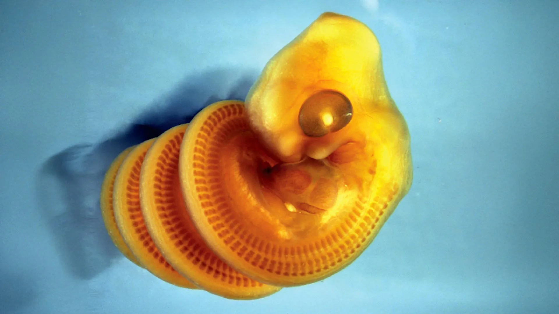
The groundbreaking findings, meticulously detailed in the prestigious journal Current Biology, illuminate a fascinating interplay of physical forces and differential growth rates at the embryonic stage. Researchers propose that the distinctive spiral form emerges as the embryo’s body undergoes rapid and extensive lengthening, a prerequisite for its adult serpentine form. Crucially, during this period of accelerated growth, the developing gut acts as a relatively fixed, internal tether. This physical constraint, a sort of internal anchor, compels the rapidly elongating body to buckle and twist into a consistent right-handed coil.
Dr. Tetsuto Miyashita, an esteemed evolutionary biologist at the Canadian Museum of Nature and the senior author and team leader of this international collaborative effort, offers an intuitive analogy to explain the phenomenon. "It’s like when you adjust the length of a strap and the longer, buckling side of the loop twists," he explains, drawing a parallel between everyday mechanics and the intricate processes unfolding within a developing snake egg. This simple yet profound mechanical principle underlies a complex biological outcome, providing a novel perspective on developmental biology.
Unraveling the Universal Mystery of Spirals in Nature
The discovery of this specific mechanism for snake embryo coiling significantly enriches our understanding of biological morphogenesis and adds a compelling new entry to the extensive and often mysterious list of naturally occurring spiral structures that continue to captivate and challenge scientists. From the grand scale of spiral galaxies to the microscopic helical structure of DNA, from the intricate geometry of a nautilus shell to the swirling patterns of hurricanes, spirals are ubiquitous in the natural world. Scientists are perpetually striving to decipher the underlying principles—be they mathematical, physical, or biological—that govern the formation of these ubiquitous and aesthetically pleasing shapes.
"There is a touch of mystery to spirals, and we are only beginning to understand how these shapes are produced in animals, such as our looping intestine, snail shells, and now these beautifully coiled snake embryos," remarks Alexandra Weber, the lead author of the study, who is now pursuing her graduate studies in zoology at the University of British Columbia. Her statement underscores the profound implications of this research, extending beyond ophidian development to a broader comprehension of biological pattern formation. The human intestine, for instance, exhibits complex coiling patterns essential for its function, and the mechanisms driving its specific morphology share conceptual parallels with the principles uncovered in snake embryos.
Miyashita further elaborates on the human fascination with spirals, noting their deep resonance in culture and art. "These puzzles beckon our curiosity. After all, spiral forms in nature have inspired human creations ranging from rotini pasta, to a barber’s pole or even portrayals of the biblical ‘Tower of Babel’," he adds. This cultural context highlights the deep-seated human appreciation for these forms, making the scientific unraveling of their origins all the more compelling. The study thus bridges the gap between fundamental biological inquiry and universal human experience.
From COVID Lockdown to a Breakthrough Discovery
The genesis of this remarkable project is itself a testament to scientific ingenuity and the ability to find opportunity in adversity. The entire research initiative blossomed out of an unusual circumstance during the unprecedented global upheaval of the COVID-19 pandemic. In 2020, with laboratories and museum collections largely inaccessible due to lockdown restrictions, Dr. Miyashita found himself working from home, grappling with the challenge of identifying a meaningful research question that his students could investigate remotely, without the need for specialized equipment or direct physical access to specimens.
"Then the lightbulb turned on. I had inherited from my PhD advisor this fascination with asymmetries in animal forms. So every time I saw images of snake embryos in papers, I wondered whether they are right- or left-handed in their coiling," Miyashita recounts. This seemingly simple observation, rooted in a long-standing academic curiosity about morphological asymmetry—a common theme in developmental biology, from the situs inversus in humans to the coiling direction of snail shells—became the unexpected cornerstone of a significant scientific endeavor. It demonstrates how foundational insights can emerge from careful observation and a persistent, inquiring mind, even under restrictive conditions. That seemingly minor question, "which way do they coil?", became the foundational hypothesis driving the entire study.
A Robust Sample: More Than 900 Snake Embryos Examined
To address this fundamental question, Miyashita enlisted the aid of Alexandra Weber, who was then an undergraduate student at Carleton University, alongside two additional undergraduate students from the University of Ottawa. Their task was to undertake a comprehensive, systematic search through a vast array of published scientific literature and accessible museum databases for photographs and illustrations of developing snake embryos.
"We obtained pictures for more than 900 embryos from 39 snake and other limbless squamate species. That’s a statistically robust sample," Miyashita emphasizes, highlighting the extensive and rigorous data collection that underpinned the study. The inclusion of nearly a thousand embryos across a diverse range of species lent considerable statistical power and broad biological relevance to their findings, ensuring that the observed patterns were not merely anecdotal but represented a widespread developmental phenomenon within limbless reptiles. This painstaking work, performed remotely, allowed the team to identify a clear and consistent pattern that had previously been overlooked or not systematically analyzed.
What emerged from this exhaustive data collection was a strikingly clear pattern. During the initial several weeks following the deposition of the eggs, the developing embryos consistently exhibited a dextral coiling pattern—meaning they spiraled to the right, as viewed from their head to their tail. This consistent orientation in early development was a critical observation.
"At these stages, the embryos don’t have muscles to move with, so different forces are making them coil right-handed," Weber explains. This crucial insight immediately ruled out active muscular contraction as the primary driver of the initial coiling. The embryos were not yet neurologically or muscularly developed enough to actively maneuver themselves into such a precise configuration. "But we didn’t know what’s making them do that," she admits, pinpointing the central mystery that the research team then set out to solve. The implication was clear: if active movement wasn’t responsible, then some inherent physical or anatomical feature of their early development must be imposing this specific twist.
The Crucial Revelation: CT Scans and a Hidden Gut Structure
A pivotal moment in the investigation arrived with the contribution of Dr. Raul Diaz, a distinguished collaborator from California State University Los Angeles. Dr. Diaz employed advanced Computed Tomography (CT) imaging technology to examine snake embryos with unprecedented anatomical detail. CT scans, which generate cross-sectional images of an object using X-rays, allowed the researchers to visualize the internal three-dimensional structure of the delicate embryos without invasive dissection, providing a non-destructive means to uncover hidden anatomical relationships.
The scans yielded a crucial and entirely unexpected revelation: an internal arrangement within the developing animal that had never been systematically documented or understood in this context. "Raul’s CT scan of a snake embryo revealed a structure we had never seen before—it was a pillar of gut stretching through the spiral of the coiling body," Miyashita exclaims, recalling the moment of discovery. He elaborates further, "There’s an intestine detached from the rest of the body, surrounded by tendrils of blood vessels from the yolk."
This observation was the missing piece of the puzzle, providing the long-sought mechanical mechanism that drove the embryonic coiling. The researchers deduced that snake embryos are evolutionarily compelled to lengthen their bodies rapidly and dramatically to achieve their characteristic elongated adult form. However, the internal organs, particularly the gut, do not grow at the same accelerated pace. This significant mismatch in growth rates—the body rapidly extending while the gut lags—creates a powerful mechanical constraint.
"So they detach the slow-growing gut, which is now tethering the lengthening body. The body buckles and twists into coiling," Miyashita meticulously explains. This ingenious developmental strategy effectively resolves the growth disparity. Furthermore, the researchers discovered that "This coiling force is directed so the embryos grow to the opposite side of the yolk. And the yolk is always to the left side of the embryo, hence the embryo will always start coiling right-handed." This elegant explanation connects the consistent left-sided position of the yolk sac—the embryo’s primary nutrient source—to the invariable initial right-handed coiling, providing a complete mechanical and spatial understanding of the phenomenon.
The Dynamic Shift: Why Coiling Direction Later Changes
The study also elucidated why this initial, mechanically imposed right-handed coiling is not a permanent state throughout the entire developmental period. As the embryos continue to mature, several factors contribute to a shift in their coiling patterns. The yolk sac, which initially dictated the coiling direction, progressively diminishes in size as its nutrients are absorbed, creating more internal space within the egg. Concurrently, the embryo’s musculature develops and strengthens, granting it the capacity for active movement and positional adjustments.
"Some remain in right-handed coils, but some recoil to the left side," Weber notes, describing the later developmental stages. "So half of these near-hatching embryos are right-handed and the other half left-handed." This observation underscores the dynamic nature of embryonic development, where initial physical constraints give way to active, muscle-driven repositioning. The research thus clearly differentiates between the earliest, passively imposed coiling and the later, actively maintained or adjusted orientations. The results conclusively suggest that the earliest direction of coiling is primarily dictated by specific developmental anatomy and intrinsic physical forces, rather than by deliberate muscular movement or genetic programming for a specific handedness.
Broader Implications: A New Model for Spirals in Biology
For Dr. Miyashita, the discovery transcends the specific case of snake embryos, illustrating a broader methodological point in scientific inquiry. It highlights how a seemingly simple observational question, pursued with diligent data collection and insightful anatomical investigation, can lead to profound biological insights. "Scientists have long been fascinated with how and why snakes evolved their strange body form. To answer that question, they tended to take a deep dive into sophisticated genetic research, looking at Hox genes, enhancers, and so on," he reflects. These genetic studies, which explore the molecular basis of limb loss and axial elongation, are undoubtedly crucial. "These are key discoveries. But here, out of the COVID lockdown, we uncovered a snake’s secret with a startlingly simple approach — just scroll through an album of snake embryos and record which way they are coiled, and take a good look at their anatomy." This underscores the value of morphology and biomechanics alongside molecular biology in understanding evolutionary adaptations.
The researchers are optimistic that the general model derived from this study—one involving differential growth rates, internal tethering, and mechanical buckling—could serve as a foundational framework for investigating other diverse spiral-shaped structures observed across the vast tapestry of living organisms. "We are now opening the possibility to develop this model further to explain other spiral forms in nature," Miyashita adds, envisioning a ripple effect of their discovery.
Weber echoes this sentiment, encapsulating the journey from curiosity to profound understanding: "This all started out with a curiosity to see if snakes are ‘handed’. It was exciting to follow it to deep insights about their evolution." This research stands as a powerful testament to the collaborative spirit of science, the unexpected avenues of discovery, and the enduring allure of fundamental biological questions. The study team represents a truly international and interdisciplinary effort, comprising dedicated scientists, students, and professors from the Canadian Museum of Nature, the University of British Columbia, Carleton University, the University of Ottawa, California State University Los Angeles, and the University of Helsinki. Their collective endeavor has not only solved a long-standing mystery in snake development but has also opened new pathways for understanding the universal language of spirals in biology.
