
 By admin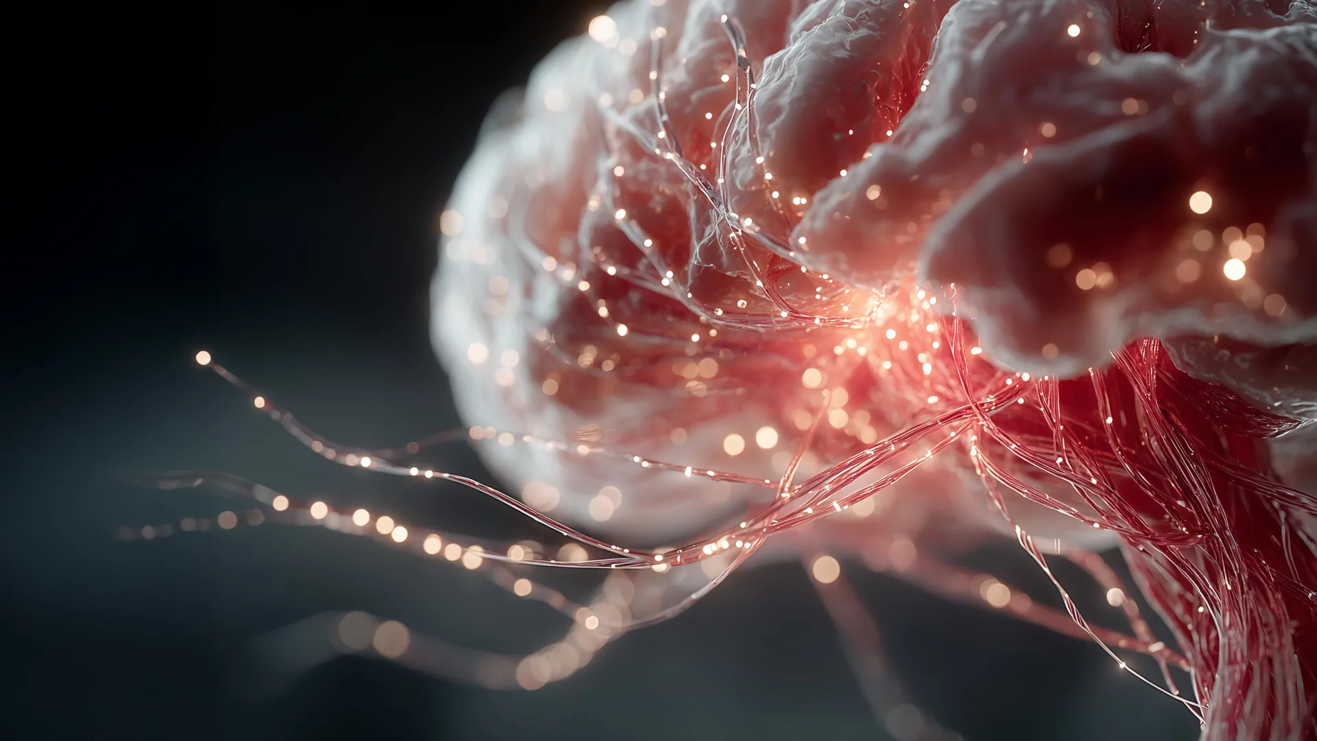
Scientists at the Mark and Mary Stevens Neuroimaging and Informatics Institute (Stevens INI) at the Keck School of Medicine of USC have uncovered compelling evidence that two neighboring types of brain tissue—gray matter and superficial white matter—may work in concert to support cognitive abilities as we age. Their groundbreaking findings suggest that the integrity of the brain’s intricate local communication pathways could profoundly influence how strongly the inevitable loss of gray matter impacts an individual’s thinking, memory, and language skills. This research offers a more nuanced understanding of brain aging, pointing towards novel avenues for fostering cognitive resilience.
Published in the esteemed journal Alzheimer’s & Dementia: The Journal of the Alzheimer’s Association, the study integrated advanced brain imaging with comprehensive cognitive testing from a cohort of 459 adults, all aged 60 and older. Critically, these participants were drawn from diverse communities across India, marking one of the first investigations into superficial white matter within a community-based population from a low- and middle-income country (LMIC). This inclusive approach is vital for overcoming the historical bias towards Western, educated, industrialized, rich, and democratic (WEIRD) populations in neuroscientific research, thereby enriching our global understanding of brain aging.
The Brain’s Intricate Architecture: Gray Matter, White Matter, and the Local Network
To truly appreciate the significance of this discovery, it’s essential to understand the fundamental components of the brain’s architecture. The human brain is a marvel of biological engineering, broadly categorized into gray matter and white matter, each playing distinct yet interconnected roles.
Gray Matter: The Processing Hub
Gray matter constitutes the outer layer of the cerebral cortex, as well as deeper nuclei within the brain. It is primarily composed of neuronal cell bodies, dendrites, unmyelinated axons, and glial cells. Often referred to as the "processing hub," gray matter is where the bulk of information processing occurs. It is responsible for a vast array of functions, including thought, language, memory, attention, perception, and voluntary movement. When we think of "brain cells," we are often picturing the neurons densely packed within the gray matter, busily computing and integrating information.
White Matter: The Communication Highways
Beneath the gray matter lies white matter, which is predominantly made up of myelinated axons – long nerve fibers that transmit signals between different gray matter regions. Myelin, a fatty sheath, insulates these axons, enabling rapid and efficient communication across vast distances within the brain. White matter tracts are often likened to the "communication highways" or "superhighways" that connect distant cities (gray matter regions), allowing for coordinated activity and complex cognitive functions.
Superficial White Matter: The Local Road Network
The Stevens INI study zeroes in on a particularly intriguing and often overlooked component of white matter: superficial white matter (SWM). This thin, delicate layer of nerve fibers lies directly beneath the gray matter covering the brain’s exterior. Unlike the long-range white matter tracts that connect distant brain regions, SWM comprises short, curved fibers that predominantly connect nearby regions of the cerebral cortex. These can be vividly imagined as the "local roads," "neural shortcuts," or "inter-regional bridges" that facilitate immediate information exchange between adjacent neighborhoods within the brain’s processing landscape.
Together, gray matter and the superficial white matter directly beneath it form a closely integrated system. If gray matter is where information is processed, superficial white matter ensures that these processing units can communicate efficiently with their immediate neighbors, fostering localized collaboration.
"Gray matter and superficial white matter are physically close and may play different roles: gray matter processes information, while superficial white matter helps nearby brain regions communicate," explained Yingxu Liu, PhD, a postdoctoral scholar at the Stevens INI and the first author of the study. "Our findings suggest that cognitive health depends not only on how much gray matter is preserved, but also on the condition of the wiring that connects it." This statement highlights a paradigm shift, urging researchers to look beyond gray matter volume alone and consider the microstructural integrity of its immediate support system.
Measuring the Brain’s Hidden Wiring: Advanced Neuroimaging Techniques
To delve into these crucial local connections, the researchers employed an advanced neuroimaging technique known as diffusion MRI (dMRI). Unlike conventional structural MRI scans that provide static images of brain anatomy, dMRI tracks the microscopic movement of water molecules through brain tissue. Water diffusion is not random in the brain; it tends to move more freely along the direction of nerve fibers and is restricted across them. By analyzing these patterns of water movement, scientists can infer the orientation, density, and integrity of nerve fibers—features that conventional brain scans cannot reveal.
The researchers specifically focused on two key measurements derived from dMRI: neurite density and the amount of freely moving water surrounding these structures. Neurites are the small projections—axons and dendrites—through which nerve cells send and receive signals. A higher neurite density generally indicates a healthier, more robust network of neural connections. Conversely, a lower density of neurites or an increase in "free water" (unrestricted water movement) can be indicative of underlying damage or disruption within the tissue. Such disruptions can be associated with various pathological processes, including the loss of myelin (demyelination), inflammation, swelling (edema), or axonal degeneration, all of which compromise the brain’s communication efficiency.
In conjunction with these detailed brain scans, participants underwent a battery of comprehensive cognitive assessments. These tests covered several critical domains of cognition, including language proficiency (comprehension, fluency, naming), memory recall and recognition, executive function (planning, problem-solving, working memory), and visuospatial ability (understanding spatial relationships). This multi-faceted approach ensured a robust and holistic evaluation of each individual’s cognitive profile.
Unveiling the Language Connection and the Role of Resilience
The study yielded several compelling insights. The most consistent and robust connection between superficial white matter health and cognitive performance was observed in the domain of language. Individuals with healthier superficial white matter, as indicated by dMRI measures, consistently tended to perform better on language tests. This association was particularly strong in the frontotemporal regions of the brain, which are known to be critically involved in complex language processes such such as recognizing words, generating fluent speech, and temporarily holding linguistic information in mind (working memory for language). This suggests that efficient local communication within these specific regions is paramount for optimal language function.
While measures of gray matter atrophy (shrinkage) remained the strongest overall predictors of cognitive ability, the researchers uncovered a critical moderating effect. They found that the impact of gray matter loss on cognition was not uniform; rather, it appeared to vary significantly depending on the health and integrity of the nearby superficial white matter.
Specifically, when these local SWM connections showed poorer integrity (indicating damage or disruption), gray matter loss was more strongly associated with impaired language performance and broader cognitive difficulties. Conversely, when superficial white matter was healthier and more intact, the link between gray matter loss and poorer cognitive outcomes was notably weaker.
This profound observation raises the exciting possibility that the condition of the brain’s local wiring could help explain a long-standing clinical puzzle: why two individuals with similar amounts of gray matter loss do not necessarily experience the same degree of cognitive decline. Some individuals appear to be more resilient to age-related brain changes than others, and this study points to SWM health as a potential key factor in that resilience.
"The findings point to superficial white matter as a possible source of resilience," affirmed Leon Aksman, PhD, assistant professor of research neurology at the Stevens INI and senior author of the study. "Two people with a similar degree of gray matter loss may not experience the same cognitive effects if the local connections surrounding that gray matter differ in health. Following participants over time will be essential to test whether preserving these connections can help maintain cognition." This concept of "cognitive reserve" or "brain reserve," where the brain’s ability to cope with pathology varies, gains a new anatomical and functional dimension with these findings.
Expanding Brain Aging Research Beyond Typical Populations
A pivotal strength of this research lies in its population source. The researchers drew their data from the Harmonized Diagnostic Assessment of Dementia for the Longitudinal Aging Study in India (LASI-DAD). This cohort is remarkably diverse, with more than half of the larger LASI-DAD population reporting low literacy levels, and approximately 60% residing in rural communities.
Populations with these socioeconomic and educational characteristics have historically been severely underrepresented in brain imaging studies, which have predominantly focused on highly educated, urban, Western cohorts. This study, therefore, represents a crucial opportunity to examine the intricate processes of cognitive aging across a significantly broader and more representative range of social, educational, and geographic backgrounds.
Intriguingly, in this specific analysis, the association between superficial white matter health and language ability was found to be even stronger among individuals who were unable to read or who read incorrectly, those who had received no formal education, and participants living in rural areas. While the researchers cautiously emphasize that these results do not establish that these social factors directly caused changes in brain tissue, they strongly suggest that the trajectory of brain aging is shaped by a complex interplay of lifelong experiences. These include educational attainment, social circumstances, overall health status, and environmental exposures, all of which contribute to an individual’s unique brain reserve and resilience. This highlights the critical need for an ecological perspective on brain health.
What Scientists Still Need to Learn: The Road Ahead
Despite its significant contributions, the study, being cross-sectional (examining participants at only one point in time), inherently has limitations. It cannot definitively establish the temporal sequence of brain changes—meaning, researchers do not yet know whether the deterioration of superficial white matter begins before gray matter loss, develops concurrently with it, or precedes measurable cognitive decline. Determining causality and progression requires longitudinal research.
Therefore, long-term studies that follow individuals as they age will be indispensable to clarify these complex relationships and understand the dynamic interplay between SWM, gray matter, and cognition over time. Future investigations will also aim to explore how other crucial biological factors, such as vascular health (blood vessel integrity), systemic inflammation, and the accumulation of Alzheimer’s disease-related proteins (like amyloid-beta and tau), interact with observed changes in both gray and white matter. This holistic approach is essential for constructing a comprehensive model of brain aging.
"A fuller understanding of brain aging requires research that reflects the world’s social, cultural, and geographic diversity," stated Arthur W. Toga, PhD, director of the Stevens INI and Provost Professor at USC. "By studying an underrepresented population and looking beyond gray matter alone, this work brings us closer to identifying the biological and social factors that may protect cognition across the lifespan."
This pioneering research from the Stevens INI fundamentally shifts our perspective on cognitive aging. By highlighting the often-overlooked role of superficial white matter and its profound influence on how gray matter loss impacts our thinking abilities, it opens new avenues for exploring biomarkers, therapeutic targets, and lifestyle interventions aimed at fostering cognitive resilience and promoting healthy brain aging for all. The emphasis on diverse populations underscores the urgent need for a global, inclusive approach to unraveling the mysteries of the aging brain.
In addition to Liu and Aksman, the study’s authors include Kirsten M. Lynch, Miguel Arce Rentería, Emma Nichols, Alden L. Gross, Lindsay C. Kobayashi, Neda Jahanshad, John P. John, Harshita V. Vishwakarma, Pranali Khobragade, Joyita Banerjee, Niranjan Khandelwal, Jyoti Dangwal, Sudhir Saxena, Nirod Medhi, Soumik Das, Prudhvinath Reddy, Pratyaksha Rana, Arjun Narula, Saravanan Kannan, Dinesh Patel, A. B. Dey, Sharmistha Dey, and Jinkook Lee.
The research was supported by the National Institute on Aging (R01AG080473, RF1AG087965, RF1AG088003, R01AG087513), the National Institute of Mental Health (R01MH134004), the National Institute of Neurological Disorders and Stroke (RF1NS136995), and the Office of the Director of the National Institutes of Health (S10OD032285).
