
 By admin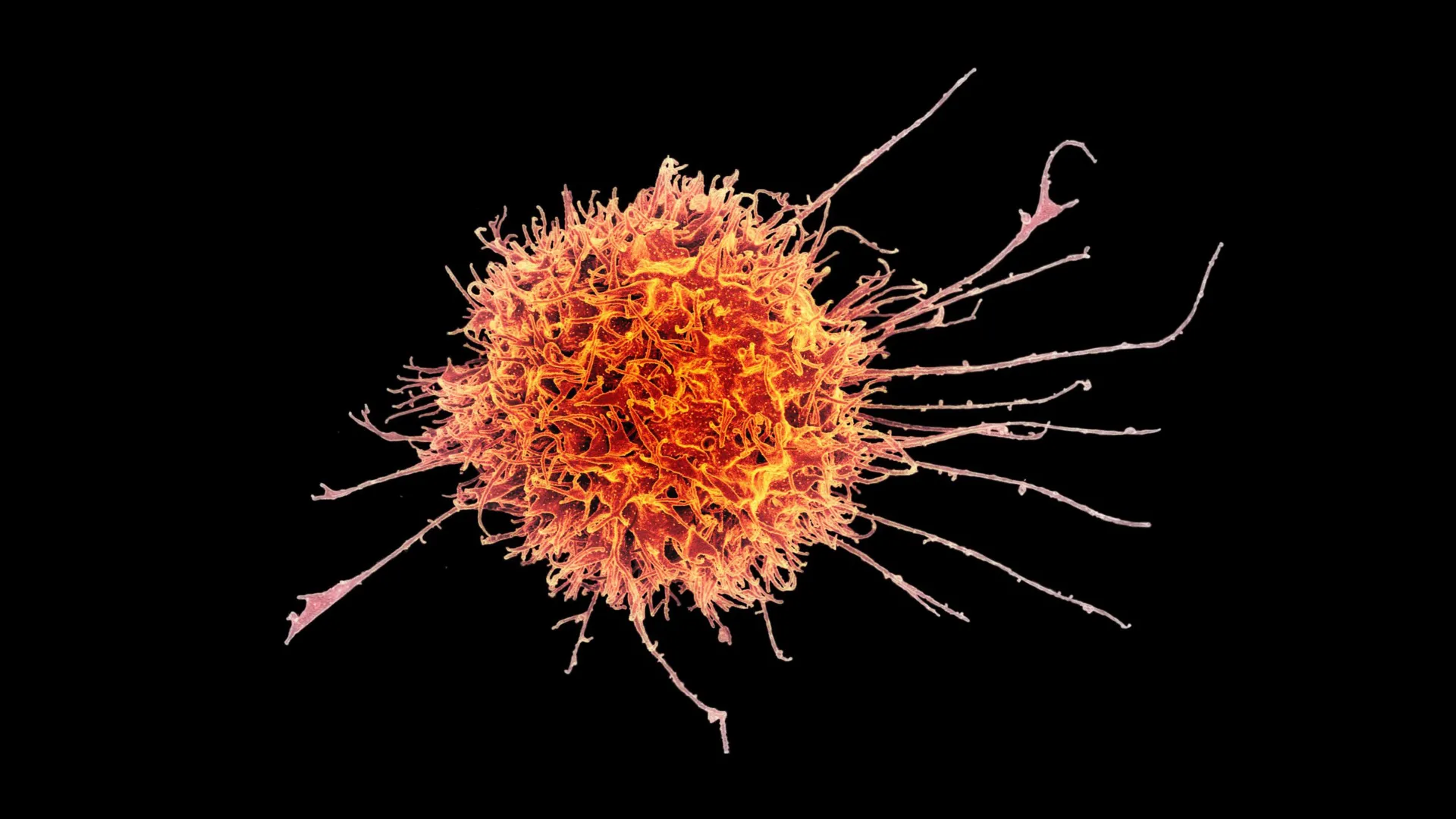
Researchers at Stanford Medicine and collaborating institutions have now developed a pioneering strategy aimed at overcoming those obstacles, offering a beacon of hope for a new generation of cancer treatments. Their innovative approach transforms natural killer (NK) cells, a powerful type of immune cell known for its innate ability to rapidly detect and destroy abnormal cells, into a specialized tissue-resident form. This modification equips these NK cells with enhanced capabilities, allowing them to effectively navigate into the challenging terrain of solid tumors and unleash their cytotoxic potential against cancer cells, a feat previously considered exceptionally difficult for conventional NK cells.
"We show that these tissue-resident natural killer cells infiltrate into the solid tumors much better than conventional natural killer cells. It was very reproducible, very striking and very clear," affirmed John Sunwoo, MD, the Edward C. and Amy H. Sewall Professor in the School of Medicine and senior author of the groundbreaking study published last month in the prestigious journal Science Translational Medicine. Dr. Sunwoo’s emphatic statement underscores the robustness of their findings, highlighting a fundamental shift in understanding how NK cells can be engineered to tackle one of oncology’s most persistent problems. This discovery could pave the way for a paradigm shift in how solid tumors are approached by cellular immunotherapies. The research represents a significant leap forward, moving beyond the historical limitations of immune cell migration and persistence within the hostile tumor microenvironment.
The meticulous work behind this discovery was a collaborative effort, with the study’s co-lead authors recognized for their instrumental contributions: Nina Horowitz, PhD, a former doctoral student in otolaryngology; Imran Mohammad, PhD, a postdoctoral fellow in the Sunwoo lab; and June Ho Shin, PhD, a senior scientist also in the Sunwoo lab. Their combined expertise in immunology, cell biology, and oncology was crucial in unraveling the complex mechanisms required to reprogram NK cells for this specialized function. This collaborative spirit, involving both seasoned researchers and emerging scientists, is a hallmark of innovative biomedical research, bringing diverse perspectives to bear on complex biological questions.
Natural Killer Cells Show Promise Against Solid Tumors: A New Frontier
The team’s experimental therapy was rigorously tested in preclinical mouse models, yielding highly encouraging results. They found that the modified natural killer cells significantly slowed the growth of several aggressive kinds of solid tumors. This effect was not only measurable but also became markedly stronger when these engineered cells were strategically paired with an antibody treatment, specifically cetuximab, which is known to help guide natural killer cells toward cancer cells by marking them for immune attack through a process called antibody-dependent cell-mediated cytotoxicity (ADCC). This synergistic approach suggests a powerful combinatorial strategy that leverages both the innate killing capacity of engineered NK cells and the precision targeting of monoclonal antibodies. The types of tumors tested included human melanoma and head and neck squamous cell carcinoma, both notoriously difficult to treat and often characterized by highly immunosuppressive microenvironments and significant metastatic potential. The ability of these modified NK cells to infiltrate and exert anti-tumor effects in these models offers compelling evidence for their potential therapeutic utility.
Beyond their direct anti-tumor efficacy, natural killer cells may also offer an important practical advantage that could revolutionize access to cell therapies. Unlike T cells, which typically require individual manufacturing from a patient’s own cells (autologous therapy) to avoid severe immune rejection, NK cells do not typically trigger a strong immune reaction when transferred from one person to another (allogeneic therapy). This critical immunological feature means that a treatment based on these modified natural killer cells could potentially be produced in large, standardized batches, frozen, and made readily available "off-the-shelf" to a wide array of patients in urgent need.
"It would be almost an off-the-shelf drug," Dr. Sunwoo explained, emphasizing the profound implications for scalability and patient access. "It could make cell therapy much more accessible to a wider variety of patients." The current logistical and financial burden associated with autologous CAR T-cell therapies—which involve apheresis, complex manufacturing, and reinfusion, often taking weeks and costing hundreds of thousands of dollars—limits their reach. An allogeneic, off-the-shelf product could drastically reduce treatment delays, manufacturing costs, and infrastructure requirements, democratizing access to cutting-edge cell therapy for a broader global patient population. This is not just a scientific breakthrough but also a potential game-changer for healthcare economics and equitable access to advanced cancer care.
Why Tissue Resident Immune Cells Matter: Unveiling Hidden Immunological Power
Natural killer cells were first identified in the 1970s, their name aptly reflecting their intrinsic ability to rapidly recognize and destroy abnormal cells, including those infected by viruses and, crucially, cancer cells. What sets NK cells apart from other key white blood cells, such as B cells and T cells, is their innate, "first-responder" nature; they do not require prior exposure to a specific target antigen to mount an attack, allowing them to respond quickly and broadly. This makes them ideal candidates for immediate action against emerging threats, distinguishing them from the adaptive immunity offered by T and B cells, which require specific antigen presentation and clonal expansion.
Historically, much of immunology has centered on immune cells circulating dynamically in the bloodstream, including B cells, T cells, and natural killer cells. These circulatory cells act as systemic patrols, constantly traveling throughout the body in search of infection and disease. However, a growing body of research, including this pivotal study, has revealed that some immune cells eventually settle inside specific tissues and undergo phenotypic and functional adaptations, taking on specialized roles tailored to their local environment. These are known as "tissue-resident" immune cells, and their importance in maintaining tissue homeostasis, mediating local immune responses, and influencing disease progression is becoming increasingly clear.
"For a long time, the study of immunology and disease in humans was concentrated on the blood immune cells," Dr. Sunwoo elaborated, highlighting a historical bias in immunological research. "With the advancement of tools and bioinformatics, we are now starting to look more at what’s going on in tissue. For most immune cells, the tissue is where the action is." This shift in focus from the bloodstream to the tissue microenvironment represents a maturation of immunological understanding. It acknowledges that the vast majority of immune interactions, whether in response to pathogens, injury, or cancer, occur not in circulation but within the complex cellular milieu of tissues. Tissue-resident cells are uniquely positioned to respond to local cues, making them potentially more effective than their circulating counterparts when engineered for site-specific tasks like tumor elimination.
Tissue-resident natural killer cells are found in various anatomical locations, including the skin, mucous membranes, lungs, and liver, where they perform organ-specific immune surveillance and effector functions. However, scientists have struggled to fully understand their exact roles and characteristics because previous studies have produced conflicting results. Some research suggested these cells were relatively weak killers and could even suppress immune activity, contributing to immune tolerance in certain contexts. In contrast, other studies found that they were highly effective at destroying target cells, displaying potent cytotoxic capabilities. This apparent contradiction underscored the need for a deeper understanding of the factors that dictate their functional plasticity.
"They may adopt different functions based on certain cues in the microenvironment and in the tissue, and differentiate into a certain kind of sub-population," Dr. Sunwoo hypothesized, acknowledging the remarkable adaptability of these cells. This plasticity means that tissue-resident NK cells are not monolithic; their behavior is profoundly influenced by the local cytokine milieu, cell-cell interactions, and metabolic environment. In some circumstances, immune-suppressing tissue-resident natural killer cells are beneficial and even essential for physiological processes. During early pregnancy, for example, specialized NK cells in the uterine lining play a crucial role in preventing the maternal immune system from attacking fetal cells and actively support placental growth, demonstrating a critical immunosuppressive function. However, for cancer treatment, the goal is precisely the opposite: to harness and amplify the more aggressive, tumor-destroying type of tissue-resident NK cell.
Finding the Right Cellular Recipe: The Goldilocks Principle
The conflicting evidence surrounding tissue-resident NK cells suggested that there were likely two or more distinct functional forms, but researchers did not fully understand how these different types developed or why their behaviors diverged so dramatically. To investigate this crucial aspect, Sunwoo’s team embarked on a systematic exploration, isolating circulating natural killer cells from healthy human blood donors and exposing them to different combinations of cellular signals and growth factors in controlled laboratory settings.
One important ingredient that emerged as central to their transformation was TGF-β (transforming growth factor beta). This ubiquitous signaling protein is produced by many cell types, including, significantly, tumor cells themselves, and plays a pleiotropic role in regulating cell growth, differentiation, and immune responses. The researchers discovered, however, that the amount and duration of the TGF-β signal were absolutely critical in determining the ultimate phenotype and function of the NK cells. This finding highlighted a sophisticated regulatory mechanism, where a single signaling molecule could elicit vastly different outcomes depending on the context of its presentation.
"It’s a Goldilocks kind of thing where if you give just enough of a TGF-β signal, then the natural killer cells become tissue resident with strong toxic activity against malignant cells," Dr. Sunwoo explained, drawing an apt analogy. "If you give too much TGF-β, they’re still tissue resident, but they’re inhibited and dysfunctional, and they don’t kill." This delicate balance is paramount. "You need it to be presented to the natural killer cells in just the right amount and in just the right manner." The experiments clearly showed that TGF-β was indeed required to initiate the transformation of circulating NK cells into a tissue-resident form. However, prolonged or excessive exposure to TGF-β, a common feature within the immunosuppressive tumor microenvironment, regrettably produced cells that were poor killers, functionally exhausted, and potentially even immune-suppressive. This explained some of the previously conflicting research and illuminated a key challenge in leveraging NK cells for cancer therapy.
A different approach, therefore, worked much better. Instead of continuous, high-dose exposure, the researchers briefly exposed natural killer cells to short-lived human epithelial tumor cells that provided a temporary, controlled burst of active TGF-β, along with other critical activating signals. This transient yet potent signaling event produced tissue-resident natural killer cells with remarkably strong tumor-killing activity. This elegant solution circumvented the immunosuppressive effects of chronic TGF-β exposure, allowing the NK cells to adopt their desired cytotoxic phenotype. Furthermore, direct physical contact with the epithelial tumor cells was also found to be essential for this transformation. Simply placing the cells nearby, without direct contact, was not sufficient to induce the aggressive tissue-resident phenotype, suggesting that additional activating signals, possibly mediated by cell-surface interactions, were involved in orchestrating this complex differentiation pathway.
"These two tissue-resident natural killer cell populations look very similar, and they have some of the same requirements, but their function seems to be on opposite ends of the spectrum," Dr. Sunwoo observed, emphasizing the subtle yet profound differences in their functional programming, driven by the nuanced presentation of microenvironmental cues.
What Makes the Strongest Killer Cells Different: Unveiling Molecular Signatures
With a reliable method established for generating the aggressive, tumor-killing tissue-resident NK cells, the team proceeded to conduct a detailed comparative analysis of the two types of tissue-resident natural killer cells: the highly cytotoxic form and the inhibited, dysfunctional form. This investigation aimed to identify the molecular markers and intracellular machinery that distinguished the potent killers from their less effective counterparts.
Both types of tissue-resident NK cells consistently displayed the surface proteins CD49a and CD103, which are recognized markers associated with tissue residency in various immune cell populations. However, a crucial distinction emerged: only the highly effective cancer-killing cells expressed CD39. CD39 is an ectonucleotidase that plays a significant role in purinergic signaling, breaking down ATP (an immune danger signal) into ADP and then AMP, ultimately leading to adenosine, which can be immunosuppressive. However, CD39 expression on NK cells in some contexts has been linked to enhanced functionality and migratory capacity, suggesting a complex role depending on the cellular context and co-expressed molecules. Its specific role here as a marker for cytotoxic tissue-resident NK cells is a novel and important finding.
Furthermore, the stronger, tumor-killing cells also contained significantly more of the molecular machinery needed to execute their cytotoxic functions. This included elevated levels of perforin, a protein that creates pores or holes in the membranes of target cells, and granzyme A, a toxic serine protease that is delivered through these perforin-created openings to induce programmed cell death (apoptosis) within the cancer cell. The increased abundance of these critical effector molecules provided a clear mechanistic explanation for their enhanced killing capabilities, confirming their potent cytotoxic programming.
Slowing Tumor Growth in Mice: Preclinical Validation
Once the researchers established a reliable method for producing the more aggressive, cytotoxic tissue-resident natural killer cells, they moved to validate their efficacy in more complex preclinical models. First, in laboratory experiments, the modified cells successfully infiltrated human tumor organoids grown in dishes, demonstrating their enhanced migratory capacity in a 3D environment that mimics the tumor architecture. This in vitro success was a crucial precursor to in vivo testing.
When injected into mice bearing human tumors, these engineered NK cells proved their mettle, significantly slowing the growth of several types of solid tumors over periods of days and weeks. These included tumors derived from human melanoma, a highly aggressive skin cancer, and head and neck squamous cell carcinoma, a devastating cancer often associated with high morbidity and mortality. The consistent anti-tumor effect across different tumor types underscored the broad applicability of this novel cellular therapy.
The strongest results, however, came when the modified natural killer cells were combined with cetuximab, a monoclonal antibody. Cetuximab targets the epidermal growth factor receptor (EGFR), which is frequently overexpressed in various cancers, including colorectal and head and neck squamous cell carcinoma. By binding to EGFR on cancer cells, cetuximab can directly inhibit tumor growth and also mark these cells for destruction by immune cells through antibody-dependent cell-mediated cytotoxicity (ADCC). Although cetuximab is approved to treat metastatic colorectal cancer and advanced head and neck squamous cell carcinoma, Dr. Sunwoo noted that it does not work especially well when used alone due to inherent tumor resistance mechanisms and the challenges of immune cell infiltration.
The combination therapy proved to be far more effective. A single dose of the modified NK cells combined with cetuximab suppressed tumor growth in mice much more effectively over one month than either treatment administered by itself. Crucially, the researchers also did not observe any apparent adverse effects or significant signs of toxicity in the treated animals, suggesting a favorable safety profile for the combination.
"Even at day 30, when the other mice were sick, the mice that received the combination seemed very healthy," Dr. Sunwoo recounted, a promising indicator of the therapy’s tolerability. However, he also wisely cautioned against extrapolating too much from mouse models to humans, adding, "This was just proof of concept." While preclinical studies are vital, human clinical trials are essential to confirm safety, efficacy, and optimal dosing in patients.
Toward an Off-the-Shelf Cell Therapy: The Road to Clinical Trials
Building on these compelling preclinical results, Dr. Sunwoo and his colleagues are now actively preparing for a Phase I clinical trial to test the combination therapy in people with advanced squamous cell carcinoma. This critical first step in human testing will primarily focus on evaluating the safety of the modified NK cells and the combination therapy, as well as determining the optimal dosage. The trial could potentially begin by the end of the year, pending crucial approval from the Food and Drug Administration (FDA), which rigorously scrutinizes all investigational new drugs and therapies.
To facilitate the rapid translation of this research into a widely accessible treatment, Dr. Sunwoo has also proactively developed and applied to patent a method for efficiently producing and expanding large numbers of these modified cells, technically known as cytotoxic tissue-resident natural killer cells. This intellectual property protection is vital for attracting investment and ensuring that the technology can be scaled for clinical and commercial use.
According to the researchers’ estimates, natural killer cells collected from a single healthy donor could potentially produce approximately 20 treatment doses in roughly two weeks. This high yield, combined with the allogeneic nature of NK cells, represents a significant logistical and economic advantage over current autologous cell therapies. "They’ll be cryopreserved, so we can make a bunch of doses and give it to different patients," Dr. Sunwoo explained, highlighting the practical benefits of an off-the-shelf product. "There would be no delay." This streamlined process could dramatically reduce the waiting time for patients, a critical factor in rapidly progressing cancers, and significantly lower the cost of manufacturing, thereby expanding access to these life-saving therapies to a much broader patient population. The promise of an immediate, readily available, and affordable cell therapy could truly transform the landscape of solid tumor treatment, offering renewed hope where options have historically been scarce.
The successful development of this innovative cell therapy is a testament to the collaborative spirit of scientific research. Researchers from Ohio State University and Washington University School of Medicine contributed to the work, underscoring the multidisciplinary effort required to tackle complex biological challenges. The study also received essential funding from the National Institutes of Health (grants R35DE030054, K22CA282364 and R25DC020174), the Tai Tsun Wu Research Fund for Natural Killer Cell Immunotherapy, and the Stanford Bio-X Fellowship. This financial and institutional support is indispensable for pushing the boundaries of biomedical science and translating laboratory discoveries into tangible clinical solutions.
