
 By admin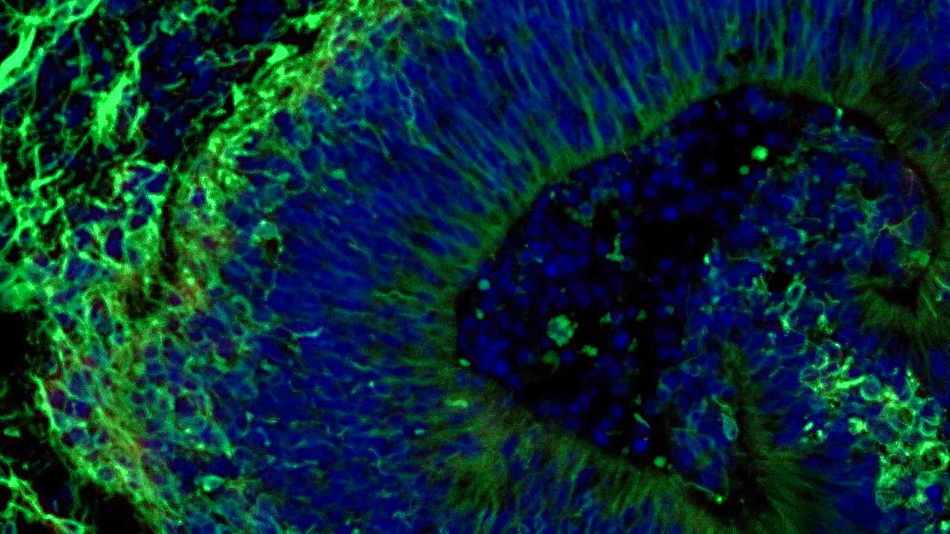
Alzheimer’s disease, the most prevalent form of dementia, currently afflicts over 7 million Americans, a number projected to rise dramatically in the coming decades. Characterized by progressive neurodegeneration, cognitive decline, and debilitating neuropsychiatric symptoms, the disease poses an immense global health challenge. Despite extensive research, a cure remains elusive, and existing treatments primarily aim to manage symptoms rather than halt or reverse disease progression. The variability in patient responses to these symptomatic treatments underscores the urgent need for personalized approaches, a gap that this Johns Hopkins study aims to bridge.
The study, partially funded by the National Institutes of Health and published in Alzheimer’s & Dementia: The Journal of the Alzheimer’s Association, not only highlights the predictive power of patient-derived organoids for drug response but also unveils the potential of tiny particles called extracellular vesicles (EVs) as novel biomarkers. These EVs, released by the organoids and carrying crucial cellular information, could offer a non-invasive means for diagnosing Alzheimer’s disease, monitoring its progression, and even assessing treatment efficacy.
Mini Brain Models: A New Frontier for Personalized Care
"Our study suggests that large-scale, patient-derived brain organoids and the vesicles they secrete can help us stage Alzheimer’s disease, investigate the mechanisms that drive it and assess how patient subgroups may respond to different treatments," states Dr. Vasiliki Machairaki, associate professor of genetic medicine at the Johns Hopkins University School of Medicine and the study’s leader. Her remarks underscore the multi-faceted utility of this technology, moving beyond simple drug screening to a comprehensive platform for understanding disease pathology and personalizing care.
The concept of "personalized medicine" is particularly critical in Alzheimer’s disease, where patients exhibit a wide spectrum of symptoms, disease progression rates, and responses to therapies. Currently, clinicians often rely on a trial-and-error approach, especially for managing neuropsychiatric symptoms (NPS) such as anxiety, depression, agitation, and psychosis, which affect nearly all Alzheimer’s patients at some stage. These symptoms significantly impact patients’ quality of life and place a substantial burden on caregivers. Selective serotonin reuptake inhibitors (SSRIs), a class of antidepressants, are frequently prescribed to manage these NPS, but their effectiveness varies widely among individuals. This variability, as Dr. Machairaki points out, is a major clinical challenge, often leading to prolonged periods of suboptimal treatment, increased side effects, and frustration for both patients and their families.
The Johns Hopkins researchers sought to address this by focusing on miniature models of the hindbrain, a crucial region located at the base of the skull responsible for regulating vital functions like breathing, sleep, and heart rate, and importantly, rich in serotonin-producing neurons. Serotonin is a key neurotransmitter targeted by SSRIs. The team’s objective was to ascertain whether these hindbrain organoids could reveal molecular signatures indicative of how patient-specific tissues might respond to escitalopram oxalate, a widely prescribed SSRI, in mitigating Alzheimer’s-associated symptoms.
Revolutionizing Research: From Patient Blood Cells to Brain Tissue
The genesis of these sophisticated brain models lies in the remarkable technology of induced pluripotent stem cells (iPSCs). The researchers initiated their work by collecting blood samples, with informed consent, from individuals diagnosed with Alzheimer’s disease at the NIH-funded Johns Hopkins Alzheimer’s Disease Research Center. These adult blood cells were then "reprogrammed" to revert to an embryonic, stem cell-like state—iPSCs. This process, pioneered by Nobel laureate Shinya Yamanaka, allows scientists to generate patient-specific cells that possess the extraordinary ability to differentiate into virtually any cell type in the human body, including highly specialized neurons.
Utilizing iPSCs derived from both Alzheimer’s patients and healthy control individuals, the team meticulously guided these cells to develop into hindbrain organoids. These pea-sized, three-dimensional clusters of brain tissue not only contained specialized serotonin-producing neurons but also exhibited a remarkable self-organizing capacity, recapitulating aspects of early human brain development. The scale of this endeavor is noteworthy; the study involved hundreds of organoids, each representing an individual patient or healthy participant. Dr. Machairaki believes this might be one of the largest brain organoid studies ever conducted in Alzheimer’s research, providing a robust platform for investigating patient-specific disease characteristics and drug responses. This large-scale approach is critical for capturing the genetic and phenotypic diversity inherent in Alzheimer’s disease.
Unveiling Alzheimer’s Molecular Footprint in Organoids
A critical validation of the organoid model’s relevance was its ability to reproduce several key biological hallmarks of Alzheimer’s disease at a molecular level. Compared to organoids derived from healthy individuals, those grown from the cells of Alzheimer’s patients displayed distinct differences in proteins crucial for inter-neuronal communication, inflammatory pathways, and other mechanisms known to be associated with the disease’s pathology. These molecular discrepancies included alterations in synaptic proteins vital for neuronal signaling and changes in proteins involved in neuroinflammation, a significant driver of neurodegeneration in Alzheimer’s. The accurate recapitulation of these disease characteristics within the organoids provides compelling evidence that these models genuinely reflect aspects of the human condition, making them invaluable tools for research.
Following this initial characterization, the researchers treated the patient-derived organoids with escitalopram oxalate. The results were illuminating and directly supported the personalized medicine hypothesis. In a subset of the Alzheimer’s patient-derived organoids, the medication elicited a noticeable increase in proteins involved in serotonin signaling and synaptic communication—the very pathways that antidepressants are designed to modulate. However, other organoids, also derived from Alzheimer’s patients, showed little to no molecular response to the drug.
This observed heterogeneity mirrors the clinical reality of varied patient responses to SSRIs in treating Alzheimer’s-related neuropsychiatric symptoms. "We used these organoids to model how some patients’ tissue may respond to a commonly prescribed SSRI," Dr. Machairaki explained. "On a large-scale level, our model may eventually be used to identify subgroups of patients, based on underlying molecular mechanisms, who are more likely to respond to certain drugs and thus help us to create precise, targeted treatments in the long run." This statement encapsulates the transformative potential of the research: moving from a reactive, empirical approach to a proactive, predictive one.
Extracellular Vesicles: A Window into Drug Response and Disease State
Beyond direct drug testing on the organoids, the team delved into the potential of extracellular vesicles (EVs) as biomarkers. EVs are nano-sized membrane-bound particles secreted by cells, acting as messengers that transport proteins, lipids, and nucleic acids between cells. Their presence in various bodily fluids, including blood and cerebrospinal fluid, makes them attractive candidates for non-invasive diagnostic and prognostic markers. The Johns Hopkins team investigated whether EVs released by the organoids could serve as indicators of Alzheimer’s disease or, crucially, help evaluate how tissue responds to treatment.
Before and after treating the organoids with escitalopram, the scientists meticulously analyzed the protein cargo within the EVs released by both patient-derived and healthy control organoids. The findings revealed that these vesicles contained a rich array of proteins integral to essential brain activities, including neuronal communication, memory formation, and neurotransmitter release.
Notably, organoids grown from the cells of people with Alzheimer’s exhibited clear alterations in several disease-associated proteins within their EVs. Specifically, levels of RAB3A, NSF, and ATCAY were significantly lower in the Alzheimer’s organoids compared to healthy controls. These proteins play pivotal roles in normal synaptic vesicle trafficking and neurotransmitter release, mechanisms often disrupted in Alzheimer’s disease.
The subsequent treatment with escitalopram led to a fascinating observation: in certain samples, levels of some EV proteins, particularly those linked to serotonin signaling and synaptic pathways targeted by antidepressants, increased after drug exposure. Similar to the direct organoid response, some organoids displayed strong molecular changes in their EV profiles, while others showed minimal or no alteration. This variation in EV response, according to Dr. Machairaki, further strengthens the hypothesis that extracellular vesicles from brain organoids could eventually provide a non-invasive "liquid biopsy" to predict which patients are most likely to benefit from a specific treatment, ushering in a new era of precision medicine for Alzheimer’s.
Paving the Way for More Realistic Brain Models and Clinical Impact
While the current study marks a significant leap forward, Dr. Machairaki acknowledges that it represents an early, albeit crucial, step. Her future plans involve developing even more sophisticated and physiologically relevant organoids. This includes incorporating immune cells, particularly microglia, which play a critical role in neuroinflammation, a hallmark of Alzheimer’s disease. Furthermore, the development of vascular-like networks within the organoids to imitate blood vessels would be transformative, allowing for better nutrient and oxygen delivery, waste removal, and modeling of the blood-brain barrier, all of which are essential for creating tissues that more closely resemble the complexity of the living human brain.
With these advancements, Dr. Machairaki envisions a future where extracellular vesicles could function as a type of liquid biopsy in clinical practice. Such a test would offer a non-invasive method to diagnose Alzheimer’s disease at earlier stages, accurately determine its progression, and even identify specific disease subtypes based on distinct molecular signatures. This would enable clinicians to select the most appropriate and personalized treatment strategies, moving away from a generalized approach to one that is tailored to the individual patient’s unique biological profile. The implications for drug development are also profound, as these organoid-EV platforms could be used to screen potential therapeutic compounds more efficiently and effectively, predicting their efficacy and identifying responders earlier in the development pipeline.
The study involved a collaborative effort among scientists, including Rachel Boyd, Daiyun Dong, Ram Sagar, Waqar Ahmed, Xenia Androni, Paul Rosenberg, Constantine Lyketsos, and Kenneth Witwer from Johns Hopkins. Additional contributions came from Anton Iliuk of Tymora Analytical Operations and Anton Porsteinsson from the University of Rochester School of Medicine and Dentistry.
This vital research received substantial funding from multiple branches of the National Institutes of Health (T32 AG058527, R01AG052510, P30AG066507, 1RF1AG083801, AGR01054771, AGR01050515, AGR01046543, and AGR01071522), alongside support from the Paul G. Allen Frontiers Foundation and the Richman Family Precision Medicine Center of Excellence in Alzheimer’s Disease at The Johns Hopkins University. The authors declared no related conflicts of interest under Johns Hopkins University policies. This collective effort underscores the multidisciplinary nature of modern scientific breakthroughs and the critical role of sustained funding in addressing complex health challenges like Alzheimer’s disease.
