
 By admin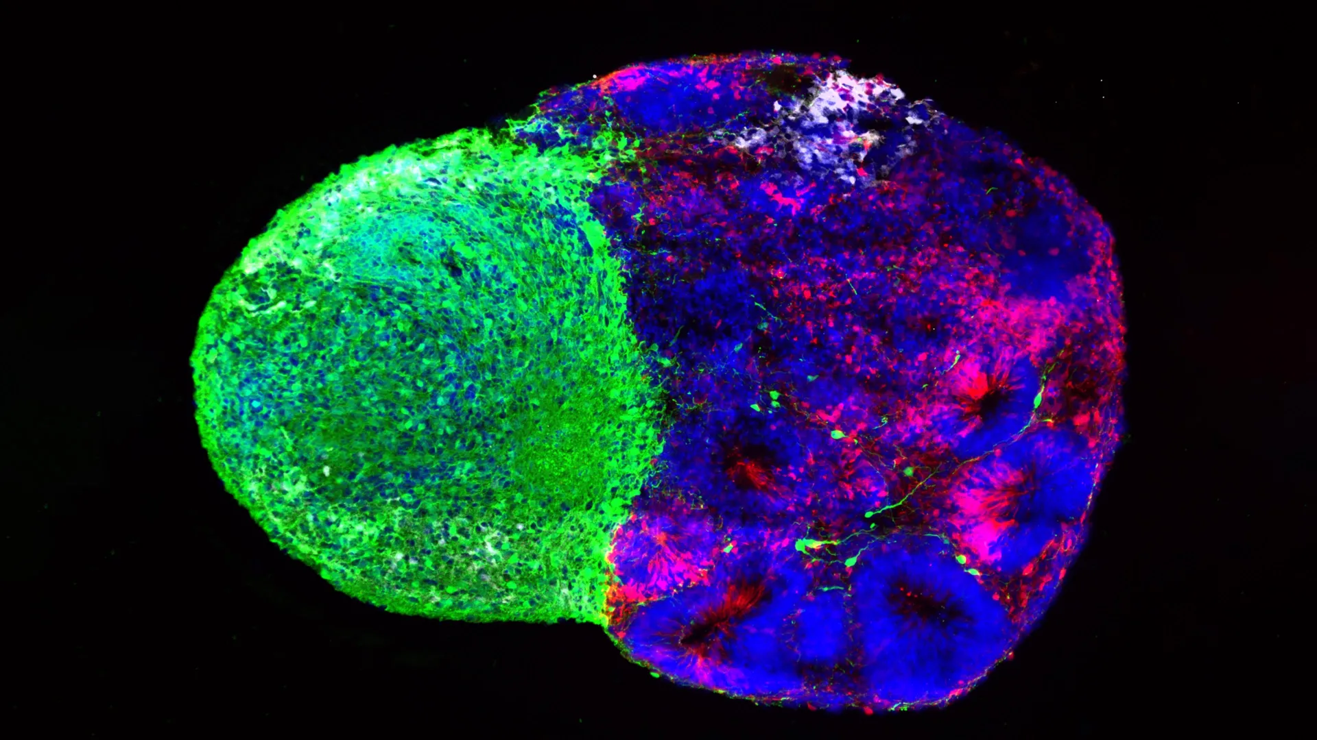
The human brain, particularly its outermost layer, the cerebral cortex, is a marvel of biological engineering. This region is the seat of our higher cognitive functions, responsible for thought, memory, language, and consciousness. The unparalleled complexity and computational power of the human cortex are directly attributable to the vast number and diverse array of neurons and support cells it contains, all largely produced by radial glia during embryonic development. Beyond sheer cell count, radial glia are also widely believed to be key contributors to the dramatic expansion of the human cortex—a characteristic that profoundly distinguishes our species from other primates and mammals. While most radial glia perform their critical developmental tasks and then disappear before birth, a perplexing and medically significant phenomenon is their re-emergence in adult brain cancers, such as glioblastoma, for reasons scientists are still striving to fully comprehend. This dual nature, essential for creation yet implicated in pathology, underscores their profound importance.
"Radial glia are the coolest cells that have ever existed," proclaims Aparna Bhaduri, an assistant professor of biological chemistry at the David Geffen School of Medicine at UCLA, her enthusiasm reflecting the scientific community’s growing appreciation for these cells. "They’re really key to making us human. But they’re also at the center of many neurodevelopmental and neuropsychiatric disorders, as well as cancer—so understanding how they make their decisions is one way to start understanding how those conditions arise." Her statement highlights the broad implications of deciphering radial glial biology, from uncovering the fundamental principles of brain formation to identifying potential therapeutic avenues for devastating diseases.
In a significant leap forward for neurodevelopmental research, two groundbreaking studies recently published in the prestigious journals Cell and Science now offer an unprecedented, closer look at the intricate mechanisms governing how radial glia make these crucial developmental choices. Bhaduri and her collaborative teams have uncovered that these powerful stem cells integrate two strikingly different, yet equally vital, categories of information: the way they process and utilize nutrients—their metabolism—and direct physical signals emanating from another critical part of the developing brain, the thalamus. Together, these convergent findings provide profound new insight into the astonishing capacity of the human cortex to produce its remarkable variety of specialized cell types, laying the groundwork for future investigations into both normal development and disease pathogenesis.
Metabolism Helps Direct Brain Stem Cells
The first study, detailed in Cell, delves into the metabolic landscape of the developing human brain, revealing an unexpected active role for cellular energy processing. This ambitious project was a collaborative effort between Bhaduri’s lab and Heather Christofk’s lab at UCLA, with co-first authors Jessenya Mil and Jose Soto leading the investigative charge. Their work sought to build a comprehensive, detailed map of metabolism within the rapidly developing human cerebral cortex.
To achieve this, the research team employed a sophisticated combination of methodologies. They analyzed precious donated human fetal tissue, providing an invaluable glimpse into in vivo development. Complementing this, they utilized brain organoids, three-dimensional cellular structures grown from human pluripotent stem cells in the laboratory, which mimic key aspects of early brain development. By integrating data from both sources, they constructed a robust metabolic atlas. The results challenged conventional wisdom, leading to a profound and unexpected conclusion: metabolism does not merely serve as a background support system, passively fueling the growth and activity of developing brain cells. Instead, it actively and directly influences which specific kinds of cells radial glia produce, fundamentally shaping the composition and function of the nascent cortex.
The researchers specifically discovered that radial glia exhibit a significant dependence on the pentose phosphate pathway (PPP), a crucial metabolic process. This pathway diverts glucose—the primary energy source for cells—to produce essential materials vital for cells undergoing rapid division. The PPP generates NADPH, a critical reducing agent necessary for synthesizing lipids and nucleotides, as well as for protecting cells from oxidative stress. It also produces ribose-5-phosphate, a fundamental building block for DNA and RNA. Given the high rates of proliferation and biosynthesis required for radial glia to generate billions of neurons and glial cells, their reliance on the PPP makes intuitive sense, providing the necessary energetic and material resources for rapid growth and differentiation.
The transformative aspect of their discovery emerged when the scientists experimentally manipulated this metabolic pathway. When they lowered the amount of available glucose or specifically interfered with the pentose phosphate pathway, the radial glia dramatically altered their developmental trajectory. Rather than proceeding with their typical program, they began generating an increased number of inhibitory neurons and other cell types that normally appear much later in development. This disruption underscores metabolism’s role not just in providing energy, but in acting as a signaling mechanism, directly influencing cell fate decisions and the precise temporal sequence of neurogenesis. An imbalance in excitatory and inhibitory neurons is a hallmark of many neurological disorders, making this finding particularly significant.
"What was surprising is that metabolism isn’t just a passive thing that happens in the background," Bhaduri reiterated, emphasizing the paradigm shift this research represents. As a member of both the UCLA Broad Stem Cell Research Center and the UCLA Health Jonsson Comprehensive Cancer Center, she bridges the gap between fundamental developmental biology and its implications for disease. "It can really control how stem cells make decisions." This revelation opens up entirely new avenues for understanding how the most fundamental cellular processes dictate complex developmental outcomes.
The immediate implications of these findings are far-reaching. The results could profoundly help scientists investigate how external factors, such as maternal nutrition during pregnancy, metabolic disorders (e.g., gestational diabetes, inborn errors of metabolism), and other environmental influences (e.g., exposure to toxins, stress) might directly affect the developing human brain. By identifying specific metabolic pathways as control points, researchers can now explore precise molecular mechanisms by which these factors contribute to neurodevelopmental conditions. Furthermore, the metabolic atlas generated by this study represents one of the most detailed and comprehensive resources yet available for researchers studying metabolism during human brain development, promising to accelerate discoveries across various fields.
A Signal Arrives Early from the Thalamus
The second groundbreaking study, published in Science and spearheaded by first author Claudia Nguyen, explored a completely different, yet equally impactful, source of developmental information. This research shifted focus from internal cellular metabolism to external inter-regional communication, specifically examining signals originating from the thalamus. The thalamus, a deep-seated structure within the brain, functions as a critical relay station, processing and transmitting sensory and motor information throughout the nervous system, including to the cerebral cortex.
For years, neuroscientists have known that neurons within the thalamus send long, intricate projections—axon fibers—that extend towards the developing cerebral cortex. These wire-like connections eventually form highly specific synaptic links with cortical neurons, establishing the crucial thalamocortical circuits that underpin sensory perception and motor control. However, detailed anatomical studies in humans have consistently shown that these thalamic projections reach the cortex remarkably early in development, long before those final, mature synaptic connections are established. This observation presented a significant conundrum, raising an important question: Why do these vital fibers arrive so prematurely if their primary role of synaptic transmission isn’t yet active?
Utilizing cutting-edge human stem cell-derived brain "assembloids," the UCLA researchers found a compelling part of the answer. Assembloids are advanced 3D culture systems that allow scientists to co-culture different brain regions, enabling the study of interactions between distinct neural tissues—a significant advantage over single-region organoids. By creating assembloids that modeled the interaction between the developing thalamus and cortex, the team observed a direct physical interaction: the thalamic projections physically touched radial glia while the brain was still undergoing its most formative developmental stages. This was not a mere passive presence but an active engagement.
This physical contact proved to be a powerful modulator of stem cell behavior. It caused the radial glia to significantly increase their production of excitatory neurons, which are the primary signal-carrying cells in the cortex, responsible for information processing and transmission. Critically, this effect was particularly pronounced for upper-layer neurons—those residing in cortical layers II and III. These upper layers are especially expanded in the human brain compared to other species and are known to be crucial for higher-order cognitive functions such as complex thought, language, and abstract reasoning. The early arrival of thalamic projections and their direct signaling to radial glia thus appear to directly contribute to the unique expansion and complexity of the human cortex.
"We already knew that these projections influence how the cortex develops," Bhaduri explained, contextualizing the discovery. "What we specifically found is that this influence comes through an actual physical connection between the projections and the radial glia—a point of contact that just hasn’t been identified before, and one that very likely does not exist in rodents." This last point is particularly salient, underscoring a potentially fundamental difference in human brain development that may explain why rodent models, while invaluable, often fail to fully recapitulate human-specific neurodevelopmental disorders. The direct, early physical interaction represents a human-specific mechanism for guiding cortical development.
A Gene Linked to Autism Enters the Picture
Further deepening their understanding, the researchers connected this vital physical interaction between thalamic projections and radial glia to a specific gene: NRXN1 (Neurexin-1). Neurexins are a family of cell adhesion molecules well-known for their critical role in the formation, stabilization, and function of synapses—the specialized junctions where neurons communicate with one another. Mutations in NRXN1 have previously been strongly associated with a range of neuropsychiatric conditions, most notably autism spectrum disorder (ASD), schizophrenia, and intellectual disability. This connection provides a powerful molecular link between a developmental process and neurological disease.
To investigate the precise role of NRXN1 in this context, the team ingeniously created assembloids derived from patient cells carrying a known NRXN1 mutation. In these disease models, the altered thalamic signals behaved differently from those observed in assembloids derived from unaffected cells. This disruption in signaling, mediated by the compromised NRXN1, profoundly shifted the delicate balance between the number of radial stem cells and the specific types and numbers of neurons they generated. The implication is clear: a subtle alteration in this early physical interaction, driven by a genetic mutation, can ripple through the developmental program, potentially leading to an imbalanced or improperly formed cortex.
This result offers researchers an invaluable window into a possible mechanism for how disturbances occurring very early in brain development—even before birth—could influence the formation and ultimately the function of the cerebral cortex, contributing to the complex etiology of disorders like autism. By providing a mechanistic pathway from a specific gene mutation to altered cellular interactions and subsequent changes in neurogenesis, this study opens up new avenues for understanding the origins of these conditions and potentially for developing early diagnostic tools or even prenatal interventions.
The Developing Brain is in Constant Communication
Although the two landmark studies focused on distinct mechanisms—one on the internal metabolic state of radial glia and the other on direct external physical signals from the thalamus—they converge on a powerful and unifying broader idea. Both investigations unequivocally demonstrate that radial glia do not make their profound developmental decisions in isolation. Instead, their behavior, their proliferation, and their differentiation into specific neuronal and glial cell types are continuously shaped and fine-tuned by a dynamic interplay of signals from their multifaceted environment. The developing brain is a hive of constant, intricate communication, where every cellular choice is influenced by a symphony of cues.
These studies also serve as a compelling testament to how dramatically organoid and assembloid technology has transformed the study of human brain development. Just a decade ago, scientists faced significant practical limitations, possessing few direct ways to investigate how uniquely human neural stem cells behave in a physiologically relevant context. Animal models, while indispensable, often fall short in recapitulating the intricate details of human-specific brain development, particularly the expansion of the cortex and the unique timing of developmental events.
Today, brain organoids and the more complex assembloids allow researchers to recreate important features of human brain development in the laboratory, offering an unprecedented platform. These sophisticated in vitro systems make it possible to address fundamental questions about human neurobiology that simply cannot be explored through animal models alone. They provide a bridge between basic cellular mechanisms and complex brain architecture, offering a powerful tool for disease modeling, drug screening, and personalized medicine.
Bhaduri expresses optimism that these pioneering findings will encourage scientists to adopt a more holistic view, recognizing metabolism and physical cellular connections not as mere background processes, but as active, potent drivers of development. This shift in perspective could unlock deeper insights into the fundamental principles governing brain formation.
"Ultimately, these studies give us a glimpse under the hood of how these cells make decisions," she concludes, emphasizing the foundational nature of the research. "Understanding those decisions is a first step toward understanding normal brain development, disease vulnerability and, potentially, how similar stem-cell programs operate in brain cancer." The ability to decode the complex language of radial glia—how they sense their environment, process information, and make fate-altering choices—holds the key to unlocking new therapeutic strategies for a spectrum of neurological and oncological challenges, ultimately advancing our understanding of what it means to be human.
This research was generously supported by a consortium of leading institutions and foundations, including the National Institutes of Health, the National Science Foundation, the Brain & Behavior Research Foundation, the Alfred P. Sloan Foundation, the Rose Hills Foundation, the Esther A. & Joseph Klingenstein Fund, the Simons Foundation, the Chan Zuckerberg Initiative, the NIH BRAIN Initiative Cell Atlas Network, the International Foundation for Ethical Research, the UCLA Broad Stem Cell Research Center’s Stem Cell Research Training Program, and the UCLA Health Jonsson Comprehensive Cancer Center and UCLA Broad Stem Cell Research Center Ablon Scholars Program. This collaborative funding underscores the widespread recognition of the profound importance and potential impact of this groundbreaking work.
