
 By admin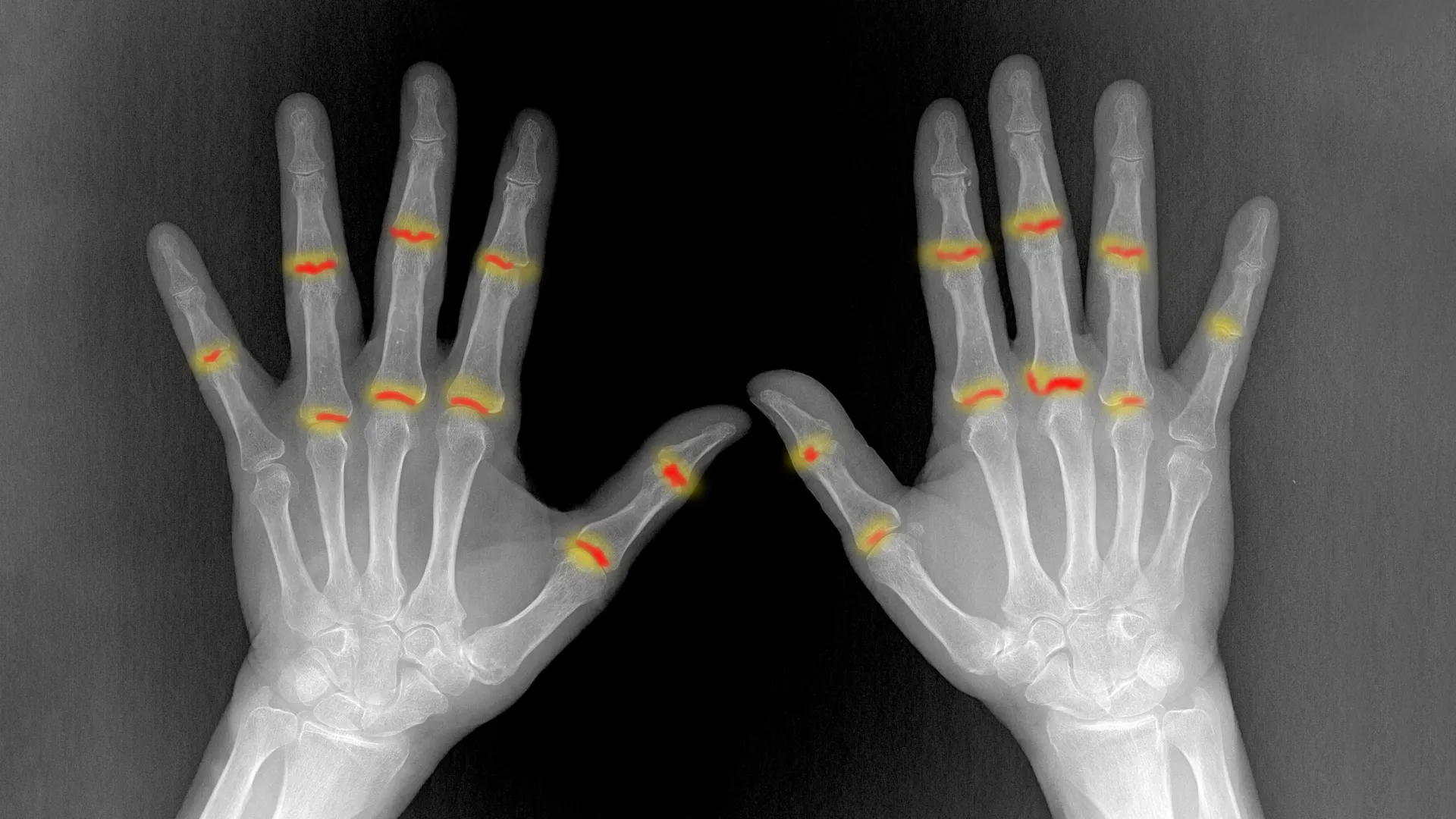
Published in the esteemed journal Nature Immunology, the study, aptly titled "The embryonic origins of site-specific arthritis," challenges conventional wisdom by proposing that the susceptibility of a joint to RA is not solely dictated by the immune system’s later assault, but also by fundamental biological differences woven into the very fabric of individual joints during their developmental stages. This paradigm shift opens new avenues for understanding, diagnosing, and potentially preventing RA.
Rheumatoid arthritis is a devastating autoimmune condition where the body’s immune system mistakenly attacks the synovium, the specialized connective tissue that lines the inner surface of joints. This misguided assault triggers a cascade of inflammation, leading to debilitating pain, swelling, and stiffness. Over time, persistent inflammation erodes cartilage, damages underlying bone, and compromises surrounding ligaments and tendons, culminating in joint deformity and severe functional disability. Globally, RA affects an estimated 1% of the adult population, with women disproportionately affected, often striking in the prime of life. Despite significant advancements in treatment, a substantial number of patients still experience disease progression and an impaired quality of life, underscoring the urgent need for a deeper understanding of its root causes.
For decades, the selective nature of RA has perplexed clinicians and scientists alike. Why do the small joints of the hands and feet bear the brunt of the disease, while others, sometimes in close proximity, remain untouched? This study meticulously investigated this phenomenon by comparing two distinct types of finger joints that exhibit vastly different patterns of disease susceptibility. The researchers focused on the proximal interphalangeal (PIP) joints, located in the middle of the fingers, which are notoriously frequent targets of RA. In stark contrast, they examined the distal interphalangeal (DIP) joints, found near the fingertips, which are generally spared from the disease’s destructive grip.
What they uncovered was remarkable: The PIP joints, those highly susceptible to RA, already possessed a larger volume of synovial tissue and contained a significantly higher concentration of PI16-positive (PI16+) fibroblasts, a specialized type of connective tissue cell, well before birth. These crucial differences, present at such an early developmental stage, suggest a compelling hypothesis: the localized tissue environment of a joint, shaped during embryonic development, may fundamentally influence its predisposition to inflammation and disease onset much later in life. This pre-programmed vulnerability could be a critical missing piece in the complex jigsaw puzzle of RA pathogenesis.
Professor Christopher Buckley, Kennedy Professor of Translational Rheumatology at the University of Oxford and a leading figure in the research, underscored the profound implications of these findings. "For decades we have known that rheumatoid arthritis selectively targets particular joints, but one of the great unanswered questions is why?" he stated. "Our findings suggest that the answer lies not only in the immune system but also in the tissues themselves. The cellular and structural characteristics established during development may help determine where inflammation takes hold later in life." This statement highlights a pivotal shift in perspective, moving beyond a purely immunological explanation to embrace the intricate interplay between innate tissue biology and immune responses. It implies that RA might not just be a disease of immune dysfunction but also a disease of developmental predispositions, where certain joints are intrinsically "primed" for inflammation.
To construct this unprecedented detailed map of human finger joints during their formative stages, the research team employed a sophisticated arsenal of cutting-edge technologies. They leveraged single-cell sequencing, a revolutionary technique that allows for the analysis of gene expression in individual cells, providing an unparalleled resolution of cellular diversity and function. This was complemented by advanced image analysis and high-resolution 3D X-ray scanning, including synchrotron-based imaging at Diamond Light Source, the UK’s national synchrotron science facility. This multi-modal approach enabled them to visualize and characterize entire joints with an astonishing level of detail, a feat often challenging to achieve with adult tissue due to the complexity and density of mature joint structures.
By observing joints during their developmental trajectory, the researchers were able to discern their fundamental composition. They found that developing joints were predominantly comprised of structural cells, such as fibroblasts and chondrocytes (cells responsible for forming cartilage), rather than immune cells, which are typically more prevalent in adult, inflamed joints. This early developmental window provided a clear view of the inherent biological blueprint of the joints. The team then meticulously examined the intricate signaling pathways and molecular cues that orchestrate the differentiation of these progenitor cells into various specialized cell types, laying the foundation for future joint function.
Of particular interest was a specific group of fibroblasts that contribute to the formation of the synovial lining. These cells are vital for joint health, producing lubricating substances like hyaluronic acid that reduce friction and facilitate smooth movement. However, in the context of arthritis, these same cells can undergo profound phenotypic changes, adopting an aggressive, pro-inflammatory, and tissue-destructive behavior. Further detailed analysis by the team suggested a fascinating developmental origin for the synovial lining: it appeared to develop from two distinct sources – cartilage and surrounding joint fibroblasts. Moreover, local microenvironmental conditions, such as areas with low oxygen levels (hypoxia), also seemed to exert a significant influence on this intricate developmental process. Understanding these precise developmental signals is crucial, as it could unlock new clues about how synovial fibroblasts acquire their diverse functions and, more importantly, how to potentially re-engineer them to restore their normal protective behavior in arthritic conditions, effectively preventing or reversing disease progression.
The researchers identified several critical differences between the RA-prone PIP joints and the generally spared DIP joints. Utilizing a specially developed image analysis tool, they definitively showed that PI16+ fibroblasts were significantly more abundant in PIP joints. These particular cells were not randomly distributed; instead, they were strategically concentrated around blood vessels, which are crucial for nutrient supply and immune cell trafficking, and at specific locations where tendons and ligaments connect to nearby bone and tissue – areas often subjected to mechanical stress and known sites of early RA inflammation.
Beyond their higher abundance and specific localization, PI16+ fibroblasts also exhibited a distinct response to inflammatory signals compared to other fibroblast populations. While both PI16+ and PI16- fibroblasts demonstrated a general pro-inflammatory reaction to external stimuli, the PI16+ cells displayed unique and pronounced changes in biological pathways directly involved in immune regulation and the organization of tissue architecture. This suggests that PI16+ fibroblasts might not just be passive recipients of inflammatory signals but active orchestrators of the inflammatory response, potentially amplifying or sustaining it in vulnerable joints.
The differences extended beyond individual cell populations, encompassing the very structure and organization of the synovial tissue itself. Employing the sophisticated high-resolution 3D imaging capabilities at Diamond Light Source, the team revealed that PIP joints possessed a greater volume of synovial tissue. Crucially, this tissue was organized in a fundamentally different manner compared to the synovium found in DIP joints. These structural variations, alongside the distinct cellular populations and their unique inflammatory responses, collectively offer a compelling explanation for why inflammation develops more readily and aggressively in some joints than in others. The altered tissue architecture could provide a more hospitable environment for immune cell infiltration and activation, further fueling the inflammatory cascade.
Dr. Sarah Davidson, a Postdoctoral Researcher at the Kennedy Institute and one of the first authors of the study, emphasized the translational potential of these findings. "We found that joints commonly affected by rheumatoid arthritis already contain distinct cellular populations before birth," she stated. "PI16+ fibroblasts were enriched in vulnerable joints and responded differently to inflammatory signals. Their location and behavior suggest they could help shape where disease develops." This underscores the idea that a "blueprint for disease" might be laid down very early in life, offering a potential window for intervention long before symptoms manifest.
The findings from this landmark study point towards a broader, more encompassing explanation for the selective nature of rheumatoid arthritis. It challenges the long-held notion that joint vulnerability is determined entirely by immune activity that occurs later in life. Instead, the propensity for inflammation to take hold may be profoundly influenced by the cellular and structural features established during the very formation of the joints in utero. In essence, each joint’s unique local biological environment, meticulously crafted during embryonic development, may pre-determine its susceptibility to rheumatoid arthritis years or even decades later.
This groundbreaking research has far-reaching implications for the future of RA research, diagnosis, and treatment. It suggests that therapeutic strategies might need to move beyond simply suppressing the immune system in adulthood. Future research could focus on understanding the precise molecular mechanisms by which PI16+ fibroblasts contribute to RA pathogenesis and exploring interventions that could modify their behavior or even target their developmental pathways. This could lead to the development of novel, highly targeted therapies that prevent the initial priming of joints for inflammation. Furthermore, identifying these early developmental markers could pave the way for predictive biomarkers, allowing for the identification of individuals at high risk for RA long before the onset of symptoms. Such early identification could enable preventative strategies or very early interventions, potentially altering the disease course dramatically and improving long-term outcomes for millions affected by this debilitating condition.
The collaborative effort behind this significant breakthrough involved scientists from the Kennedy Institute of Rheumatology, University of Oxford, working in conjunction with colleagues from the University of Birmingham, University College London, and the advanced imaging capabilities of Diamond Light Source. The study was generously supported by the Medical Research Council (MRC), underscoring the importance of sustained investment in fundamental research to unravel complex diseases like rheumatoid arthritis. This collaborative, multi-institutional approach exemplifies the power of interdisciplinary science in addressing some of the most challenging questions in human health.
