
 By admin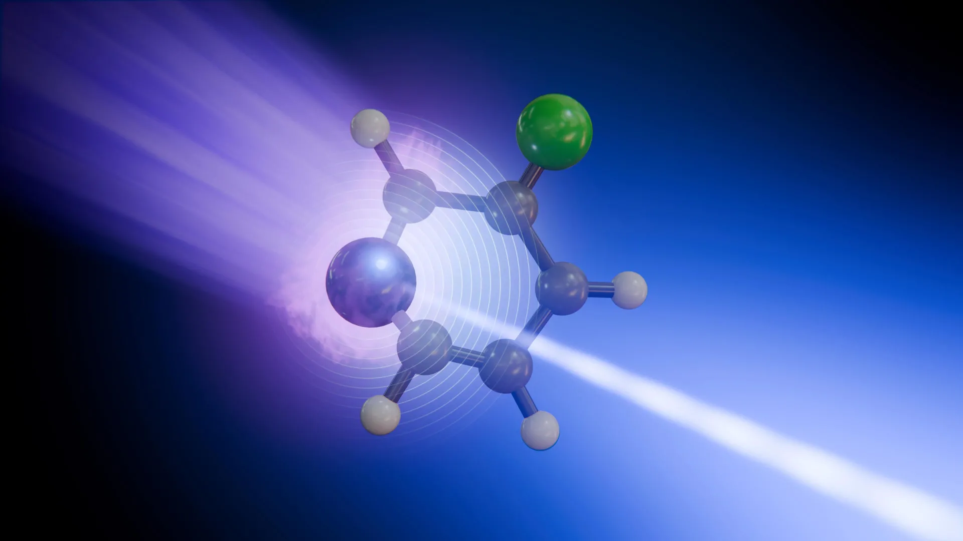
For decades, scientists have strived to unravel the intricate mechanisms governing photochemistry – the study of chemical reactions initiated by light. These processes often unfold on femtosecond (quadrillionths of a second) and picosecond (trillionths of a second) timescales, making direct observation a formidable challenge. Traditional spectroscopic methods typically provide an averaged view of molecular changes, obscuring the unique contributions of individual atoms within a complex molecular structure. This new research shatters that limitation, offering a high-resolution, atom-specific perspective on how absorbed light energy is converted and propagated.
The European XFEL: A Stroboscope for Molecules
The key to this observational breakthrough lies in the European XFEL (X-ray Free-Electron Laser), located in Hamburg, Germany. As the world’s largest X-ray laser, it generates ultrashort, extremely bright X-ray flashes that are powerful enough to probe the innermost electronic shells of atoms. These X-ray pulses act like an atomic-scale stroboscope, allowing researchers to capture snapshots of molecular transformations with femtosecond precision. By varying the delay between an initial light pulse that triggers a reaction and the subsequent X-ray flash that probes it, scientists can assemble a molecular "movie" of events unfolding in real time.
The specific technique employed was time-resolved X-ray photoelectron spectroscopy (tr-XPS), carried out at the Small Quantum Systems (SQS) instrument of European XFEL. Tr-XPS is particularly powerful because it measures the kinetic energy of electrons ejected from a molecule after it’s hit by an X-ray photon. Crucially, the binding energy of these deeply bound "core" electrons is highly sensitive to the local chemical environment around the atom they originate from. This means that as a molecule undergoes structural changes or redistributes electron density following light absorption, the core electron binding energies of its constituent atoms will subtly shift, providing a unique fingerprint of the changes occurring at each specific atomic site.
Deciphering the Molecular Dance of 3-fluoropyridine
The researchers chose 3-fluoropyridine, a relatively small, ring-shaped organic molecule, as their experimental subject. Its compact size and the presence of both nitrogen and fluorine atoms made it an ideal candidate for studying site-specific energy flow. When 3-fluoropyridine absorbs a brief ultraviolet (UV) laser pulse, its electrons are kicked into a higher energy state – a process known as electronic excitation. This surge of energy doesn’t just sit statically; it immediately triggers a cascade of events.
The molecule, typically flat in its ground state, quickly distorts and bends out of shape. This structural rearrangement is not random; it’s a critical step towards energy dissipation. As the molecule changes geometry, it passes through what scientists call a "conical intersection." These intersections are short-lived but incredibly important regions on a molecule’s potential energy surface where two electronic states become degenerate, meaning their energies become equal. At these critical junctures, the movements of electrons and the atomic nuclei (the "cores" of the atoms) become strongly coupled.
Conical intersections act like "funnels" or "chutes," enabling ultrafast non-adiabatic transitions. This means the molecule can rapidly switch from an excited electronic state back to its ground electronic state without emitting light (a process called non-radiative decay). Instead, the excess electronic energy is efficiently converted into vibrational energy – the jiggling and shaking of the atoms within the molecular framework. This rapid energy conversion is a fundamental process in many light-driven phenomena, including photostability in biological molecules and the initial steps of light harvesting in solar energy conversion. Without these efficient funnels, excited molecules would linger in higher energy states, potentially leading to harmful chemical reactions or inefficient energy utilization.
After navigating the conical intersection, 3-fluoropyridine returns to its ground electronic state, but now carries a substantial amount of vibrational energy. This energy then propagates through the molecular structure as vibrations, causing the atoms to oscillate and relax back to their equilibrium positions. It is this subsequent redistribution of vibrational energy that the scientists were able to meticulously track, atom by atom.
Different Atoms, Different Stories: The Atomic-Specific Readout
One of the most profound discoveries of the study was that different atoms within the same molecule reported distinct aspects of this complex energy transformation. The fluorine atom, located at a specific position on the pyridine ring, served as a relatively straightforward indicator. Its signal primarily reflected how the molecule’s overall vibrations relaxed over time. As the excess energy dissipated through the molecule’s structural motions, the fluorine atom’s core electron binding energy shifted in a way that clearly mapped this vibrational relaxation process. It acted as a passive, yet precise, sensor for the global structural dynamics.
The nitrogen atom, on the other hand, told a more intricate story. Being an integral part of the pyridine ring and playing a more direct role in the initial electronic excitation, its core electron signal was far more complex. It reflected not only the changing structure of the molecule but also the transient redistribution of electron density immediately following light absorption and during the passage through the conical intersection. The nitrogen atom, therefore, offered a richer, more nuanced perspective, capturing both the electronic and structural facets of the transformation.
"We can now see that not every atomic site tells the same story in the signals we capture from our X-ray pulses," stated Antonio Picón from the Instituto de Ciencia de Materiales de Madrid Consejo Superior de Investigaciones Científicas (ICMM-CSIC), a co-author of the study. His insight underscores the revolutionary nature of this atom-specific approach. "Some atoms report where the charge is going, while others reveal how the whole molecule vibrates." This ability to differentiate between electronic and vibrational energy flow at specific atomic locations is a critical advancement, moving beyond the averaged data typically obtained in ultrafast spectroscopy. It provides an unprecedented level of detail, allowing researchers to pinpoint the exact roles individual atoms play in the energy dissipation pathway.
Reconstructing a Picosecond Transformation: A Symphony of Experiment and Theory
To piece together this molecular saga, the research team employed a meticulous experimental protocol. First, an ultrashort UV laser pulse delivered energy to a cloud of 3-fluoropyridine molecules, initiating the photochemical process. Then, after precisely controlled delays ranging from femtoseconds to picoseconds, a soft X-ray pulse from the European XFEL was fired. This X-ray pulse ionized the molecules by removing deeply bound electrons, specifically targeting either the nitrogen or fluorine atoms depending on the X-ray energy tuned. The energies of these ejected electrons were then measured with high precision.
By repeating this measurement many times, with the X-ray pulse arriving at incrementally different delays after the initial laser pulse, the researchers were able to reconstruct a detailed timeline of how the chemical environment surrounding each atom changed over just a couple of picoseconds – an astonishingly short duration. Each data point contributed to building a comprehensive picture of the molecular evolution, capturing the fleeting moments of electron excitation, structural distortion, and subsequent energy redistribution.
However, raw experimental data alone often cannot fully explain the complex quantum mechanical phenomena at play. This is where advanced computer simulations and theoretical models became indispensable. The experimental signals, rich in information but inherently complex, were interpreted and validated using sophisticated computational chemistry techniques. These simulations provided a theoretical framework to connect the observed shifts in core electron binding energies to the underlying electronic state changes, bond length alterations, and vibrational modes within the molecule. This synergistic approach, combining cutting-edge experimental measurements with robust theoretical modeling, was crucial for extracting the deep insights into the molecular dynamics.
A New Window on Ultrafast Photochemistry and Its Vast Implications
The findings of this study are far-reaching, demonstrating the unparalleled capability of ultrashort, high-brightness X-ray pulses from facilities like the European XFEL to disentangle some of the fastest and most interconnected motions in matter. While 3-fluoropyridine is a relatively simple molecule, the methodology established here is highly versatile and can be applied to increasingly complex systems.
The potential applications are immense:
- DNA Photostability: Understanding how DNA withstands constant exposure to UV light is crucial for comprehending mutagenesis and cancer prevention. DNA’s remarkable ability to dissipate harmful UV energy rapidly, often through conical intersections, prevents damage. This technique could provide atom-specific insights into these protective mechanisms.
- Light-Harvesting Materials: The efficiency of solar cells and artificial photosynthesis devices hinges on how quickly and effectively absorbed light energy can be channeled and converted. By tracking energy flow at the atomic level in novel materials, scientists can design more efficient light harvesters.
- Functional Organic Molecules: Many organic molecules are designed to react to light for applications ranging from drug delivery to advanced sensors and displays. A detailed understanding of their photochemistry is essential for optimizing their performance.
- Biomolecular Building Blocks: Proteins, enzymes, and other complex biomolecules undergo light-driven processes. This technique could shed light on their dynamic behavior and functions.
"This is what European XFEL was built to enable: watching chemical change where it begins, at specific atomic sites and on its natural timescale," remarked Daniel Rivas, a former instrument scientist and now guest scientist at SQS, and a co-author of the study. His statement encapsulates the transformative potential of this research. "By combining multi-site sensitivity with femtosecond resolution, we are opening a new window on the microscopic mechanisms that govern photochemistry."
In essence, this work represents a paradigm shift in how scientists can study light-induced chemical reactions. By moving beyond a bulk or averaged view and peering into the individual atomic contributions to molecular transformations, researchers are gaining an unprecedented level of mechanistic understanding. This new atomic-scale cinematography of energy flow promises to accelerate discovery in fields from fundamental chemistry and biology to materials science and renewable energy, ultimately paving the way for innovations inspired by nature’s own mastery of light.
